
Percutaneous nephroscope lithotripsy and lithotripsy training model and preparation method thereof

A training model and nephroscopic technology, applied in the medical field, can solve the problems of lack of lesions, small convenience, and small skill coverage, achieve simple training environment requirements, improve surgical skills, and achieve routine effects

Pending Publication Date: 2022-03-01
BEIJING BOYI AGE MEDICAL TREATMENT TECH CO LTD
View PDF6 Cites 1 Cited by
  • Summary
  • Abstract
  • Description
  • Claims
  • Application Information

AI Technical Summary

Problems solved by technology

[0003] The current training methods for doctors are as follows: 1) The use of dry modules (bean clamping, ferrule, perforation, paper-cutting, silicone imitation meat, etc.) can only train basic abilities such as spatial sense conversion, hand-eye coordination, and hand coordination, and the skill coverage is small. , cannot meet the needs of human surgical operations (such as the use of energy devices and surgical procedures, etc.);
[0004] 2) Although a single isolated organ allows doctors to train real touch and energy equipment, it lacks lesions and distortion, and can only allow doctors to practice individual techniques such as suturing and cutting;
[0005] 3) Although the 3D simulator is relatively convenient, the human tissue is a complex multi-layer material, and because its properties depend on the number of layers, the thickness of the layers, the direction of muscle fibers, humidity, temperature, etc., the tactile feel of the 3D simulation is always Failure to meet clinical needs;
[0006] 4) Animal surgery requires supporting conditions such as animal operating room, temporary animal breeding room, and anesthesia equipment. The investment cost of venues, personnel, and equipment is high, and the convenience is small. It does not form the operability of daily training. In addition, it is like isolated pig kidneys. It cannot provide doctors with the environment of perirenal fat wrapping and the pathological state of renal cysts;

Method used


Image

  • Percutaneous nephroscope lithotripsy and lithotripsy training model and preparation method thereof

Examples


preparation example Construction

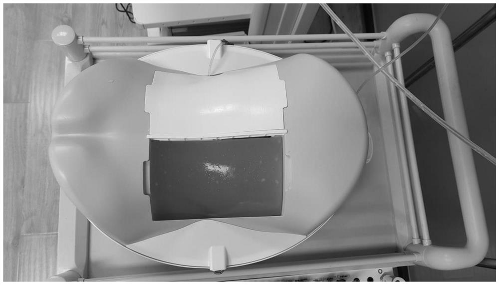
[0063] In a second aspect, the present invention provides a method for preparing a percutaneous nephrolithotomy training model, comprising the following steps:

[0064] a. Open the kidney, place a stone in the calices, and close the kidney;

[0065] b. Wrap the kidney obtained in step a with simulated fat, and place it in a container;

[0066] c. Filling the container described in step b with an ultrasound-permeable material to prepare a percutaneous nephrolithotripsy training model.

[0067] It should be noted that the angle at which the kidney is placed in the container can be placed with reference to the position of the kidney in the human body, for example, the kidney is placed in the container at an angle of 30 degrees.

[0068] The preparation method of the percutaneous nephrolithotomy lithotripsy training model provided by the present invention is simple and quick, convenient for obtaining materials, and low in cost.

[0069] In some preferred embodiments, the means o...

Embodiment 1

[0073] (1) Choose the pig's kidney with ureter length 2cm;

[0074] (2) Choose stones with a diameter of 0.5-0.8cm, which can be irregular;

[0075] (3) Open a 1 cm incision on the dorsal side of the renal pelvis, and place stones with a diameter of 0.5-0.8 cm into the upper, middle and lower calyces respectively;

[0076] (4) Suture the dorsal incision of the renal pelvis;

[0077] (5) Bind the opening end of the end of the ureter to the perfusion head and perfusion tube, and the perfusion length is 5cm;

[0078] (6) Chicken oil, uniform in thickness (thinner, about 7mm);

[0079] (7) Wrap the chicken fat completely on the surface of the kidney, and use a medical coupling agent to ensure a tight fit between the kidney and the chicken fat;

[0080] (8) Take 25g of agar and 1000g of water (ratio 1:40), boil and let stand to 70°C;

[0081] (9) Tilt the kidney by 30° and place it in a 20*30*20 box;

[0082] (10) Introduce the agar after standing still in the box that kidney ...

Embodiment 2

[0085] (1) Select sheep's kidney with ureter length 3cm;

[0086] (2) Choose stones with a diameter of 0.5-2cm, which can be irregular;

[0087] (3) Open an incision of 2.5 cm on the dorsal side of the renal pelvis, and place stones with a diameter of 0.5-2 cm into the upper, middle, and lower calyces;

[0088] (4) Adhesive closure of the dorsal incision of the renal pelvis;

[0089] (5) Bind the opening end of the end of the ureter to the perfusion head and perfusion tube, and the perfusion length is 8cm;

[0090] (6) Suet, uniform in thickness (thinner, about 5mm);

[0091] (7) Wrap the suet completely on the surface of the kidney, and use a medical coupling agent to ensure the tight fit between the kidney and the suet;

[0092] (8) Take 1500ml of A and B silica gels, mix and stir evenly, and let stand;

[0093] (9) Tilt the kidney by 30° and place it in a 20*30*20 box;

[0094] (10) Import the static silica gel into the box containing the kidney, let it stand for 4-6 h...


PUM

PropertyMeasurementUnit
Thicknessaaaaaaaaaa
Particle sizeaaaaaaaaaa
Diameteraaaaaaaaaa

Abstract

The invention provides a percutaneous nephroscope lithotripsy and lithotripsy training model and a preparation method thereof, and relates to the technical field of medical treatment. The percutaneous nephroscope lithotripsy and lithotomy training model comprises a container, an ultrasonic permeable material, simulated fat and a kidney, the container is filled with the ultrasonic permeable material, the kidney is wrapped by the simulated fat, the simulated fat is fixed in the ultrasonic permeable material, the simulated fat comprises chicken fat, leaf fat or animal fat, and the kidney is fixed in the simulated fat. The perirenal fat texture and perirenal substance filling can be fully simulated; and stones are fixed in the kidney calyx of the kidney. The training model is convenient to take materials, fully simulates touch and force feedback of real operation, and can be used for training of kidney ultrasound image recognition, puncture, expansion, lithotripsy and the like, so that medical staff can obtain real skill training touch, and surgical skills are improved; the training model is simple in training environment requirement, saves investment of sites, labor and the like, and realizes daily operation training.

Description

technical field [0001] The invention relates to the field of medical technology, in particular to a percutaneous nephrolithotripsy training model and a preparation method thereof. Background technique [0002] With the rapid development of contemporary science and technology, medical technology is changing with each passing day. However, most surgeons still use the teaching mode of "teaching by master and apprentice" and "training with surgery". In order to provide doctors with sufficient opportunities to accumulate skills and reduce the risk of practicing medicine. Surgery has extremely high requirements for doctors' operational proficiency. If doctors want to acquire effective skill accumulation, they need a lot of realistic training. [0003] The current training methods for doctors are as follows: 1) The use of dry modules (bean clamping, ferrule, perforation, paper-cutting, silicone imitation meat, etc.) can only train basic abilities such as spatial sense conversion, ...

Claims


Application Information

Patent Timeline
IPC IPC(8): G09B9/00G09B23/28
CPCG09B9/00G09B23/28
Inventor 李微
Owner BEIJING BOYI AGE MEDICAL TREATMENT TECH CO LTD
Learn More