Application of alpha-ketoglutaric acid in preparation of medicine for treating myocardial infarction
A technique for myocardial infarction and ketoglutaric acid, which is applied in the field of biomedicine and can solve the problems of α-ketoglutaric acid affecting myocardial cell proliferation and the like
- Summary
- Abstract
- Description
- Claims
- Application Information
AI Technical Summary
Problems solved by technology
Method used
Image
Examples
Embodiment 1
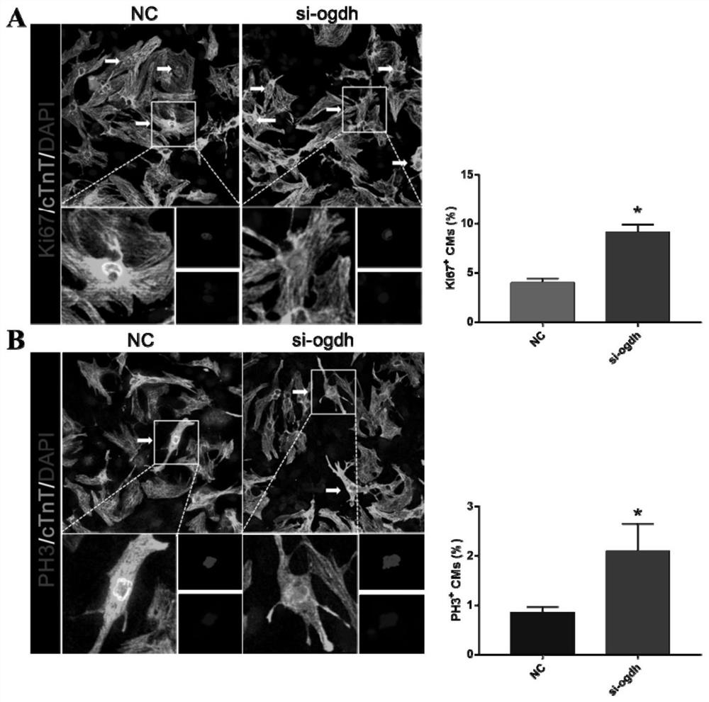
[0034] α-ketoglutarate can promote the proliferation of primary cardiomyocytes cultured in vitro
[0035] The cardiomyocytes of neonatal SD rats were extracted, and the specific method was as follows:
[0036] (1) Prepare 10 ml of type II collagenase and trypsin solutions with concentrations of 0.08% and 0.125%, respectively.
[0037] (2) The ventricle tissue of suckling mice was cut out by ophthalmologist, and stored in pre-cooled Hanks buffer.
[0038] (3) Shred the tissue to 1mm 3 Small pieces, then rinse again with pre-cooled Hanks buffer solution.
[0039] (4) Suck off the washing solution, add an appropriate amount of trypsin, gently pipette repeatedly for 1 minute, and discard the liquid after the tissues have gathered into a ball.
[0040] (5) Add collagenase, blow off, and digest at 37°C for 30 minutes.
[0041] (6) Gently tap and beat repeatedly, and after the tissue block dissolves, add 1 ml of fetal bovine serum to terminate digestion.
[0042] (7) Filter the ...
Embodiment 2
[0049] α-ketoglutarate can promote the proliferation of cardiomyocytes in developing mice
[0050] The 7-day-old mice were divided into the experimental group and the control group, and received intraperitoneal injection of α-ketoglutarate and PBS every day, and the heart weight ratio was evaluated after 2 weeks, and further detected by wheat germ agglutinin (WGA) staining Cardiomyocyte size. The results showed that injection of α-ketoglutaric acid could increase the heart weight ratio of mice ( Figure 4 ), but the volume of cardiomyocytes did not change significantly ( Figure 5 ). The expression of proliferation markers (EdU pulse labeling) was detected by immunofluorescent staining, such as Figure 6 As shown, α-ketoglutarate injection can promote the proliferation of cardiomyocytes in mice.
Embodiment 3
[0052] α-Ketoglutarate improves post-infarction cardiac function and infarct size and promotes proliferation of adult cardiomyocytes
[0053] (1) To construct a mouse model of myocardial infarction, divide the mice into the experimental group and the control group, and receive daily intraperitoneal injection of α-ketoglutarate and PBS after myocardial infarction. After continuous injection for 2 weeks, two-dimensional echocardiography Cardiac function detected by graph (EF%, FS%, LVIDs and LVIDd at 4th week after operation); Masson staining was used to detect myocardial infarct size (4th week after operation); expression of proliferation markers was detected by immunofluorescent staining (EdU pulse marker ). The results showed that α-ketoglutarate could improve cardiac function ( Figure 7 ), reducing the infarct size ( Figure 8 ), promote the proliferation of adult cardiomyocytes ( Figure 9 ).
PUM
 Abstract
Description
Claims
Application Information
 - Generate Ideas
- Intellectual Property
- Life Sciences
- Materials
- Tech Scout
- Unparalleled Data Quality
- Higher Quality Content
- 60% Fewer Hallucinations
Browse by: Latest US Patents, China's latest patents, Technical Efficacy Thesaurus, Application Domain, Technology Topic, Popular Technical Reports.
© 2025 PatSnap. All rights reserved.Legal|Privacy policy|Modern Slavery Act Transparency Statement|Sitemap|About US| Contact US: help@patsnap.com


