Acromioclavicular joint CT image processing method

A technology of CT images and acromioclavicular joints, applied in the field of CT images, to achieve good matching and reduce the effect of acromion impingement

Pending Publication Date: 2021-12-07
ANHUI PROVINCIAL HOSPITAL
View PDF0 Cites 1 Cited by
  • Summary
  • Abstract
  • Description
  • Claims
  • Application Information

AI Technical Summary

Problems solved by technology

However, no scholars have used CT to measure the acromioclavicular joint, and CT cannot be routinely used during surgery to judge the vertical and horizontal displacement of the acromioclavicular joint and whether the placement of the clavicular hook plate is reasonable.

Method used


Image

  • Acromioclavicular joint CT image processing method
  • Acromioclavicular joint CT image processing method
  • Acromioclavicular joint CT image processing method

Examples


Embodiment Construction

[0036] The present invention is described in further detail now in conjunction with embodiment.

[0037] A kind of CT image processing method of acromioclavicular joint, comprises the steps:

[0038] (1) Obtain CT data of patients

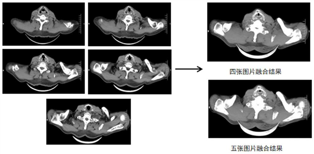
[0039] (2) Select CT images with acromioclavicular joints for image fusion

[0040] First, from the patient's CT scan images, a suitable image with the acromioclavicular joint is selected for image fusion. The selection of CT images is very important. The selected images need to show the cross-sectional images of the acromioclavicular joint, and at the same time, there should not be too many interference parts, otherwise, the fused images will also have interference parts, which will affect the follow-up. Positioning of the clavicle hook. Such as figure 1 Shown is the process of fusing images of the left acromioclavicular joint of the patient. It is advisable to select the first four cross-sectional CT images of this patient. The addition of the ...


PUM


Abstract

The invention belongs to an acromioclavicular joint CT image processing method. The method comprises the following steps: (1) acquiring CT data of a patient; (2) selecting a CT image with an acromioclavicular joint part for image fusion; and (3) post-processing the fused image to obtain an image of the acromioclavicular joint. The CT image processing method for the acromioclavicular joint aims to research the corresponding relation between the normal distal clavicle end and the acromion, and provides a reliable basis for reduction of dislocation of the acromioclavicular joint and fracture of the distal clavicle end; and, in addition, the method can enable a clavicle hook steel plate to be placed at the optimal position of the acromioclavicular joint, enable the prebending plasticity of the clavicle hook steel plate to optimally adapt to the form of acromion, achieve better matching, and reduce complications such as acromion collision and dissolution.

Description

technical field [0001] The invention belongs to the technical field of CT images, and in particular relates to a method for processing CT images of acromioclavicular joints. Background technique [0002] Acromioclavicular joint dislocation is a common injury of the shoulder, accounting for about 40% of shoulder joint injuries in athletes, and the incidence of associated related injuries is 18%-39.3%, including rotator cuff injuries accounting for about 32.3% and cartilage injuries accounting for about 30.6% %, labral injury accounted for 22.6%. [0003] X-ray examination is a low-radiation dose and economical examination method. It is still a routine method for detecting dislocation of the acromioclavicular joint in clinical practice in quite a few hospitals, and it is of great significance for the follow-up of the acromioclavicular joint and the guidance of functional exercise. . Conventional X-ray diagnosis of acromioclavicular joint dislocation includes the following me...

Claims


Application Information

Patent Timeline
IPC IPC(8): G06T5/50G06T5/00G16H30/20
CPCG06T5/50G16H30/20G06T2207/10081G06T2207/20221G06T2207/30008G06T5/70
Inventor 夏睿
Owner ANHUI PROVINCIAL HOSPITAL
Why Eureka
Try Eureka