
Head fixing device for anesthetized rats in small animal PET-CT scanning

A technology of PET-CT and fixation device, which is applied in the direction of radiological diagnostic equipment, computerized tomography scanner, application, etc. It can solve the problems of distortion of scanning results and difficult observation of mouse brain structure, so as to ensure stability and improve convenience sex, to avoid skewed effects

Pending Publication Date: 2021-11-05
XUZHOU MEDICAL UNIV
View PDF0 Cites 0 Cited by
  • Summary
  • Abstract
  • Description
  • Claims
  • Application Information

AI Technical Summary

Problems solved by technology

[0003] When the traditional small animal PET-CT scans an anesthetized rat, the whole head of the rat lies on the plane of the scanning bed, so the scanned image of the rat is different from the image shown in the rat brain atlas, and the scan It can be observed that the rat brain is in a downward and forward posture as a whole, so the scanned image does not match the anatomical image of the rat brain anatomy atlas. The side view of the rat brain shown in the rat brain anatomy atlas is Upward and the uppermost part is horizontal, if the scanned image is adjusted by software, it will affect some structures of the scanned image, which will easily cause distortion of the scanning result, and it is not easy to observe the corresponding shortcomings of the mouse brain structure

Method used


Image

  • Head fixing device for anesthetized rats in small animal PET-CT scanning
  • Head fixing device for anesthetized rats in small animal PET-CT scanning
  • Head fixing device for anesthetized rats in small animal PET-CT scanning

Examples


Embodiment 1

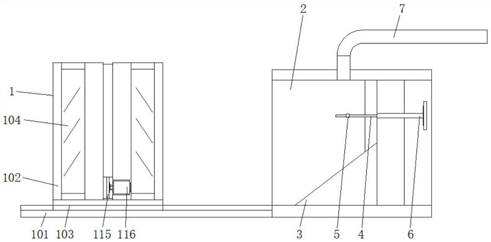
[0026] Such as figure 1 As shown, this embodiment proposes a head immobilization device for small animal PET-CT scanning anesthetized rats, including an auxiliary clamping limit mechanism 1 and a plastic cover 2, and the plastic cover 2 communicates with the anesthetic gas tube 7 , the anesthesia gas tube 7 can introduce the anesthesia gas into the inside of the plastic cover shell 2, so that the anesthesia gas can provide anesthesia treatment to the experimental mice, the plastic cover shell 2 is provided with a fixed base 3, and the fixed base 3 has an inclined triangular structure, which can be used for experiments. The mouse provides a place to place it, and a locking screw 6 is also pierced in the plastic cover shell 2, and the end of the locking screw 6 is connected with a backguy 4. and be fixed on the collar 5, the incisors of the experimental rat can be inserted into the collar 5, and the backguy 4 can be pulled, so that the collar 5 cooperates with the backguy 4 to f...

Embodiment 2

[0028] The solution in Embodiment 1 will be further introduced in combination with specific working methods below, see the following description for details:

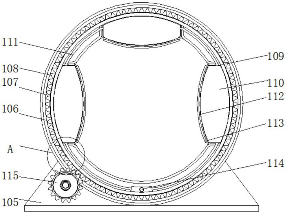
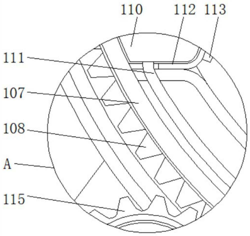
[0029] Such as figure 1 , figure 2 with image 3As shown, as a preferred embodiment, on the basis of the above method, further, the auxiliary clamping and limiting mechanism 1 includes a slot plate 101, a casing 102, a fixed slider 103, a transparent partition 104, a limiting base 105, Guide inner groove 106, ring groove plate 107, teeth 108, cavity 109, air bag 110, connecting air pipe 111, flexible pressure plate 112, rubber spacer 113, gas injection valve 114, adjustment gear 115 and servo motor 116, groove plate 101 Fixedly connected with the fixed base 3, through the fixing of the slot plate 101 and the fixed base 3, the auxiliary clamping and limiting mechanism 1 can be connected with the plastic cover shell 2, so as to improve the overall convenience of the device for anesthetizing the experimental mice. The s...


PUM


Abstract

The invention discloses a head fixing device for anesthetized rats in small animal PET-CT scanning, and relates to the technical field of scanning anaesthesia in rats, in particular to a head fixing device for anesthetized rats in small animal PET-CT scanning. The head fixing device comprises an auxiliary clamping limiting mechanism and a plastic cover shell. The plastic cover shell is internally provided with a fixed base. A locking screw is further arranged in the plastic cover shell in a penetrating mode, and the end of the locking screw is connected with a pull wire. A visible channel can be effectively provided for a housing through a transparent partition plate, so that the state of an experimental rat in the housing can be observed conveniently. A guide inner groove in the housing can effectively limit the rotation range of an annular groove plate and can effectively guarantee the rotation stability of the annular groove plate. The servo motor can push the annular groove plate to rotate in the housing by adjusting the cooperation between a gear and teeth, thereby effectively changing the rotation angle of the annular groove plate, adjusting the annular groove plate to a proper position, and turning over the experimental rat for scanning experiments.

Description

technical field [0001] The invention relates to the technical field of scanning anesthetized rats, in particular to a head fixation device for small animal PET-CT scanning anesthetized rats. Background technique [0002] The full name of PET is positron emission computed tomography, which is an imaging device that reflects the gene, molecule, metabolism and functional status of lesions. It uses positron nuclide-labeled glucose and other human metabolites as imaging agents, and reflects its metabolic changes through the uptake of imaging agents by lesions, thereby providing clinical biological metabolism information of diseases. It is a new milestone in the development of life science and medical imaging technology. [0003] When the traditional small animal PET-CT scans an anesthetized rat, the whole head of the rat lies on the plane of the scanning bed, so the scanned image of the rat is different from the image shown in the rat brain atlas, and the scan It can be observe...

Claims


Application Information

Patent Timeline
IPC IPC(8): A61B6/03A61B6/04A61B6/00A61D7/04
CPCA61B6/037A61B6/0421A61B6/508A61D7/04
Inventor 王德广徐垚唐广胜
Owner XUZHOU MEDICAL UNIV
Learn More