Method for decolorizing transparent large-volume sample and acquiring three-dimensional data online

A three-dimensional data, large-volume technology, applied in the field of biomedical optical imaging, can solve problems such as difficulties in implementing in vivo perfusion, slowing down the process of data acquisition, and slow efficiency of soaking and decolorization

Pending Publication Date: 2021-09-14
HUST SUZHOU INST FOR BRAINMATICS
View PDF5 Cites 0 Cited by
  • Summary
  • Abstract
  • Description
  • Claims
  • Application Information

AI Technical Summary

Problems solved by technology

However, for large animals such as rhesus monkeys and domestic pigs, it is very difficult to implement in vivo perfusion. Therefore, there is often a lot of blood left in the biological tissues obtained from large animals, and the efficiency of overall soaking and decolorization is very slow. Slow down the process of getting data

Method used


Image

  • Method for decolorizing transparent large-volume sample and acquiring three-dimensional data online
  • Method for decolorizing transparent large-volume sample and acquiring three-dimensional data online
  • Method for decolorizing transparent large-volume sample and acquiring three-dimensional data online

Examples


Embodiment 1

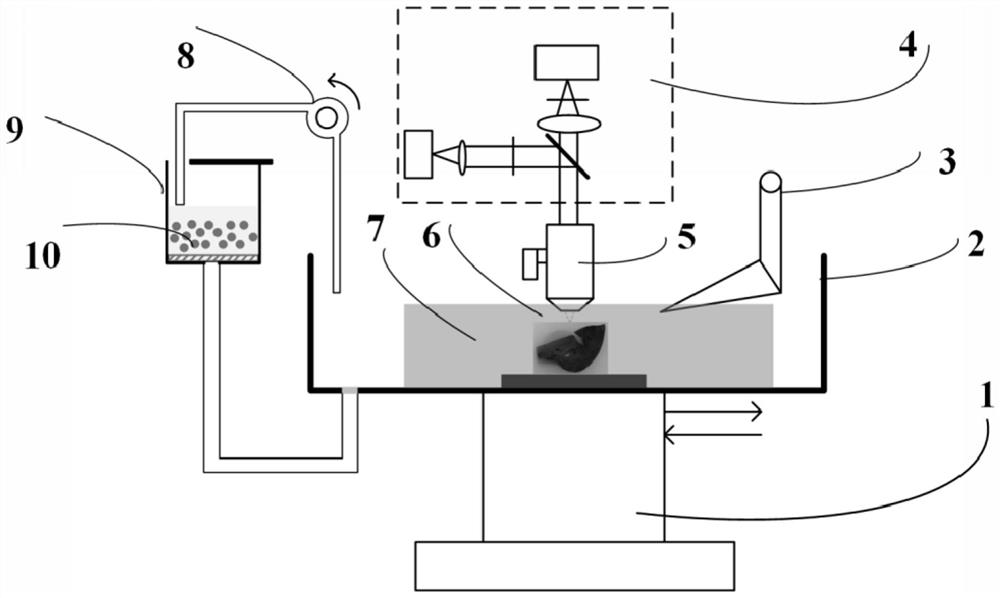
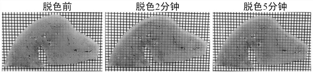
[0073] The imaging solution in Example 1 is composed of a concentration of 30wt% fructose, 20wt% urea and 20v / v% dimethyl sulfoxide, 5V / V% N-methyldiethanolamine and 0.5wt% 3-[3-(cholamide Propyl) dimethylamino] propanesulfonic acid inner salt aqueous solution. The biological sample tissue is the liver tissue of an adult domestic pig without perfusion, and the three-dimensional data acquisition is completed according to the following steps:

[0074] S1, first obtain the liver tissue of an adult domestic pig as a biological sample tissue, soak it in 4% paraformaldehyde solution and fix it for 3 months until the sample becomes hard.

[0075] S2, performing pretreatment on the biological sample tissue, that is, immersing the biological sample tissue in an imaging solution for at least 24 hours, so as to first dehydrate and decolorize the surface.

[0076] S3, performing agarose embedding on the pretreated biological sample tissue.

[0077] S4, the embedded biological sample tis...

Embodiment 2

[0088] The imaging solution in Example 1 consists of an aqueous solution with a concentration of 30wt% fructose, 20wt% urea, 20v / v% dimethyl sulfoxide, and 10v / v% N-methyldiethanolamine. The biological sample tissue is the liver tissue of an adult domestic pig without perfusion, and the three-dimensional data acquisition is completed according to the following steps:

[0089] S1, first obtain the liver tissue of an adult domestic pig as a biological sample tissue, soak it in 4% paraformaldehyde solution and fix it for 3 months until the sample becomes hard.

[0090] S2, performing pretreatment on the biological sample tissue, that is, immersing the biological sample tissue in an imaging solution for at least 24 hours, so as to first dehydrate and decolorize the surface.

[0091] S3, performing agarose embedding on the pretreated biological sample tissue.

[0092] S4, the embedded biological sample tissue is soaked in the transparent solution again for not less than 24 hours. ...


PUM


Abstract

The invention discloses a method for decolorizing a transparent large-volume sample and acquiring three-dimensional data online, which comprises an embedding step, two immersion steps before and after the embedding step, and a cyclic optical imaging and slicing step, wherein an imaging solution in the immersion steps at least comprises sugar or alcohol or a sugar alcohol compound, urea, and an amino alcohol compound. The optical transparency and microsection tomography technologies are combined and alternately used, and the imaging solution performs real-time optical transparency operation on the sample, balances the refractive index of the tissue sample, eluates the heme in the sample to remove the influence of the heme on optical imaging, improves the depth of single imaging, reduces the number of microsection times of the sample, and improves the acquisition efficiency of the three-dimensional data.

Description

technical field [0001] The invention relates to the technical field of biomedical optical imaging, in particular to a method for online decolorization of transparent large-volume samples and acquisition of three-dimensional data. Background technique [0002] The composition of living organisms is complex and diverse, and its structure and function have always been the problems explored by scientists. Obtaining high-resolution three-dimensional structural information of biological tissues at the level of organs and even the whole body is very important for us to deeply understand and solve many biological and medical problems. Optical imaging technology can obtain three-dimensional data of biological sample cell resolution, but due to the Opacity leads to serious scattering and absorption of light signals, which not only affects the imaging quality, but also affects the time of three-dimensional imaging of the sample. [0003] Tissue light transparency technology solves the...

Claims


Application Information

Patent Timeline
IPC IPC(8): G01N1/30G01N1/36G01N1/28G01N1/06G01N21/84G01B11/24
CPCG01N1/30G01N1/36G01N1/286G01N1/06G01N21/84G01B11/24G01N2001/2873
Inventor 杨孝全张云飞李向宁龚辉
Owner HUST SUZHOU INST FOR BRAINMATICS
Why Eureka
Try Eureka