Artery intracavity ultrasound imaging method, artery intracavity ultrasound imaging device, equipment and artery intracavity ultrasound imaging system

An ultrasonic imaging method and intracavity ultrasonic technology, applied in the electronic field, can solve problems such as large interval distance, multiple blood vessels and lesion information, and "sawtooth" phenomenon, so as to improve accuracy, improve diagnosis and treatment effect, and avoid image distortion.

Active Publication Date: 2021-07-16
SONOSCAPE MEDICAL CORP
View PDF10 Cites 0 Cited by
  • Summary
  • Abstract
  • Description
  • Claims
  • Application Information

AI Technical Summary

Problems solved by technology

Although coronary angiography and other vascular imaging techniques are considered the "gold standard" for evaluating coronary artery disease, there are still certain limitations. For example, angiography cannot make a functional judgment of stenosis, and (angiography) imaging only shows the lumen Therefore, we need a "third eye"—IVUS (intravascular ultrasound) and other intravascular ultrasound imaging techniques to see through arteries (such as coronary arteries) and obtain intra-arterial images. Understanding the situation in the arterial lumen and evaluating its physiological characteristics, so as to choose a reasonable treatment strategy, provide more important information for optimizing stent implantation and reducing stent-related problems in clinical practice
[0004] At present, the retraction of intra-arterial ultrasound imaging includes two modes: slow and fast. Slow retraction means longer imaging time. At the same time, with the contraction and relaxation of the heart, repeated imaging and "sawtooth" phenomenon will appear in ultrasound imaging; Fast retraction means that the retraction distance of the imaging probe per unit time is large, resulting in too large a distance between each frame of imaging, and more blood vessel and lesion information will be lost, which further leads to the problem of inaccurate measurement of the length of arterial lesions, which in turn leads to The selection of the length of the implanted stent is inaccurate, and the arterial interventional treatment cannot be accurately guided

Method used


Image

  • Artery intracavity ultrasound imaging method, artery intracavity ultrasound imaging device, equipment and artery intracavity ultrasound imaging system
  • Artery intracavity ultrasound imaging method, artery intracavity ultrasound imaging device, equipment and artery intracavity ultrasound imaging system
  • Artery intracavity ultrasound imaging method, artery intracavity ultrasound imaging device, equipment and artery intracavity ultrasound imaging system

Examples


Embodiment 1

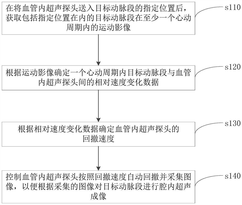
[0066] Please refer to figure 1 , figure 1 The flow chart of the intraarterial ultrasound imaging method provided in this embodiment; the method mainly includes:

[0067] Step s110, after sending the intravascular ultrasound probe (IVUS transducer) into the designated position of the target artery segment, acquiring motion images of the target artery segment including the designated position within at least one cardiac cycle;

[0068] The moving image of the target artery segment may refer to a static image, that is, a multi-frame image collected in a continuous time, or may be a dynamic image, which is not limited here.

[0069] In addition, in this embodiment, there is no limitation on the image category of the moving image. Optionally, the moving image may specifically be a digital subtraction angiography image. The process of moving images within a cycle is specifically: acquiring a digital subtraction angiography image of a target artery segment including a designated p...

Embodiment 2

[0106] Please refer to Figure 8 , Figure 8 A structural block diagram of an intraarterial ultrasound imaging device provided for this embodiment; the device may include: an image acquisition unit 110 , a relative velocity analysis unit 120 , a retraction velocity setting unit 130 , and a probe retraction acquisition unit 140 . The intraarterial ultrasound imaging device provided in this embodiment can be compared with the above-mentioned intraarterial ultrasound imaging method.

[0107] Wherein, the image acquisition unit 110 is mainly used to acquire the motion image of the target artery segment including the specified position within at least one cardiac cycle after sending the intravascular ultrasound probe into the designated position of the target artery segment;

[0108] The relative velocity analysis unit 120 is mainly used to determine the relative velocity change data between the target artery segment and the intravascular ultrasound probe within a cardiac cycle ac...

Embodiment 3

[0112] Please refer to Figure 9 , Figure 9 A structural block diagram of a computer device provided in this embodiment; the device may include: a memory 300 and a processor 310 . For computer equipment, refer to the introduction of the above-mentioned intraarterial ultrasound imaging method.

[0113] Wherein, the memory 300 is mainly used for storing programs;

[0114] The processor 310 is mainly used to implement the steps of the above-mentioned intraarterial ultrasound imaging method when executing the program.

[0115] Please refer to Figure 10 , is a schematic structural diagram of a computer device provided in this embodiment. The computer device may have relatively large differences due to different configurations or performances, and may include one or more processors (central processing units, CPU) 322 (for example, one or one processor) and memory 332, one or more storage media 330 (such as one or more mass storage devices) for storing application programs 342 ...


PUM


Abstract

The invention discloses an artery intracavity ultrasonic imaging method. The withdrawing speed of a probe is determined according to relative speed change data between a target artery section and an ultrasonic probe when the ultrasonic probe is static, so that the influence of the movement process of the artery cavity along with the heartbeat on the withdrawing image acquisition process of the probe can be reduced; loss of useful data can be avoided while repeated imaging is avoided; an appropriate number of arterial cavity images are enabled to be acquired; the occurrence of image distortion is avoided; the accuracy of withdrawing image acquisition is improved, so that a reliable length value is provided for an arterial intervention doctor in the arterial intervention treatment process, and the accuracy of the arterial lesion length measured according to artery intracavity ultrasonic imaging is improved; and the diagnosis and treatment effect can be effectively improved. The invention also discloses an artery intracavity ultrasonic imaging device, computer equipment, a computer readable storage medium and an artery intracavity ultrasonic imaging system, which have the beneficial effects.

Description

technical field [0001] The present invention relates to the field of electronic technology, in particular to an arterial intracavity ultrasonic imaging method and device, a computer device, a computer-readable storage medium and an arterial intraluminal ultrasonic imaging system. Background technique [0002] Cardiovascular disease is the number one health killer in the world, and it is also the main cause of death among Chinese residents. Among them, coronary heart disease is one of the diseases with the highest mortality and disability rates, and it is also the disease with the fastest growing mortality rate in my country. [0003] Vascular imaging techniques such as coronary angiography can evaluate vessel size, plaque characteristics, and stent implantation effects, etc. The size of the lumen and lesion characteristics can be evaluated before surgery, and the postoperative intraluminal imaging enables the operator to evaluate the stent at the stent level The effect of im...

Claims


Application Information

Patent Timeline
Patent Type & Authority Applications(China)
IPC IPC(8): A61B8/08
CPCA61B8/0891A61B8/481
Inventor 张勇秦志飞孙银君田秋芳朱彦聪
Owner SONOSCAPE MEDICAL CORP
Why Eureka
Try Eureka