DCNN-based cancer full-view digital pathological section survival analysis method

A technology of digital pathology slides and survival analysis, applied in the field of intelligent medical image processing, can solve problems such as difficulty for doctors to pay attention to details, large WSI size, and patient analysis

Active Publication Date: 2021-05-18
FUDAN UNIV
View PDF7 Cites 4 Cited by
  • Summary
  • Abstract
  • Description
  • Claims
  • Application Information

AI Technical Summary

Problems solved by technology

Although professional doctors can complete the diagnosis by analyzing WSI, it is difficult for doctors to pay attention to all the details due to the huge size of WSI
At the same time, there are many factors that affect the prognosis of cancer, and it is difficult for doctors to analyze the relevant information of the patient's prognosis through pathological sections.

Method used


Image

  • DCNN-based cancer full-view digital pathological section survival analysis method
  • DCNN-based cancer full-view digital pathological section survival analysis method
  • DCNN-based cancer full-view digital pathological section survival analysis method

Examples


Embodiment Construction

[0029] The embodiments of the present invention will be described in detail below, but the protection scope of the present invention is not limited to the examples.

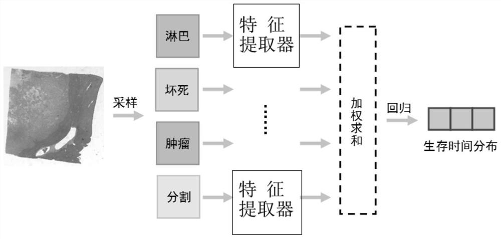
[0030] use figure 1 In the framework of the process, 1169 pathological slices with known distribution of lymphatic dense areas, necrotic areas, tumor areas, and paracancerous areas of 363 patients were used for training to obtain a prognosis model that can automatically perform automatic survival analysis on cancer pathological slices.

[0031] The specific process is:

[0032] (1) Before training, one image with a length and width of 1024 pixels was randomly sampled from the lymphatic dense area and the necrosis area; five images with a length and width of 1024 pixels were randomly sampled from the tumor area. Moreover, the masks of the above four regions were combined into a 4-channel image, and scaled to a length and width of 512 pixels as a global segmentation map of pathological slices. The 7 sampled image...


PUM


Abstract

The invention belongs to the technical field of medical image intelligent processing, and particularly relates to a DCNN-based cancer full-view digital pathological section survival analysis method. The method comprises the following steps: respectively taking a plurality of image blocks from a lymphatic dense region, a necrosis region and a tumor region in a pathological section, sending the image blocks and global segmentation of the section into a convolutional neural network, and acquiring survival time distribution of the section through forward propagation The death probability of the patient in a plurality of time intervals can be obtained from the predicted survival time distribution, and the survival probability of the patient before a plurality of time nodes can be obtained through accumulation. Local images of the sections and global tissue distribution serve as input, the survival time distribution of the patient serves as output, a doctor is helped to estimate the prognosis condition of the patient, and therefore early clinical diagnosis of cancer is assisted.

Description

technical field [0001] The invention belongs to the technical field of medical image intelligent processing, and in particular relates to a method for survival analysis of pathological slices, and more specifically, relates to a method for survival analysis of cancer full-view digital pathological slices based on a convolutional neural network. Background technique [0002] With the development of whole slide scanning technology, a large number of tissue slices are scanned as whole-field digital pathological slides (Wholeslide image, WSI), stored in digital form, and widely used in cancer pathological diagnosis. Although professional doctors can complete the diagnosis by analyzing WSI, it is difficult for doctors to pay attention to all the details of WSI due to its huge size. At the same time, there are many factors that affect the prognosis of cancer, and it is difficult for doctors to analyze the relevant information of the patient's prognosis through pathological slides....

Claims


Application Information

Patent Timeline
Patent Type & Authority Applications(China)
IPC IPC(8): G06T7/00G06T7/11G06N3/04G06N3/08
CPCG06T7/0012G06T7/11G06N3/08G06T2207/20081G06T2207/20084G06T2207/30096G06N3/047
Inventor 高强颜波谭伟敏丁光宇凌宇
Owner FUDAN UNIV
Why Eureka
Try Eureka