Near-infrared fluorescent parathyroid gland recognition enhancement method based on AR technology

A parathyroid, AR technology, applied in the field of near-infrared fluorescent parathyroid identification enhancement

Active Publication Date: 2021-04-16
THE FIRST AFFILIATED HOSPITAL ZHEJIANG UNIV COLLEGE OF MEDICINE
View PDF13 Cites 1 Cited by
  • Summary
  • Abstract
  • Description
  • Claims
  • Application Information

AI Technical Summary

Problems solved by technology

[0011] After our investigation, we found that there is no relevant research report on the application of near-infrared autofluorescence detection technology and AR technology in parathyroid detection at home and abroad.

Method used


Image

  • Near-infrared fluorescent parathyroid gland recognition enhancement method based on AR technology
  • Near-infrared fluorescent parathyroid gland recognition enhancement method based on AR technology
  • Near-infrared fluorescent parathyroid gland recognition enhancement method based on AR technology

Examples


Embodiment 1

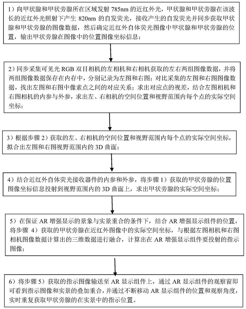
[0091] Embodiment 1 A method for identifying and enhancing near-infrared fluorescent parathyroid glands based on AR technology described in the present invention includes the following steps:

[0092] 1) Emit 785nm near-infrared light to the area where the thyroid gland and parathyroid glands are located. The thyroid gland and parathyroid glands produce 820nm autofluorescence under the irradiation of near-infrared light of this wavelength, receive the generated autofluorescence and simultaneously acquire the thyroid gland and parathyroid glands Then determine the position of the thyroid gland and parathyroid gland in the near-infrared autofluorescence image, and output the image coordinate information of the position of the parathyroid gland in the image;

[0093] 2) Synchronously collect the left and right sets of image data obtained by the left and right cameras of the visible light RGB binocular camera, and store the two sets of image data in the memory, and record them as t...

Embodiment 2

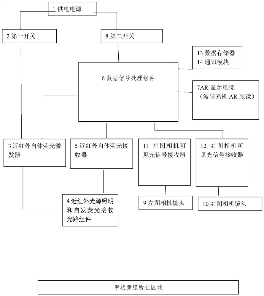
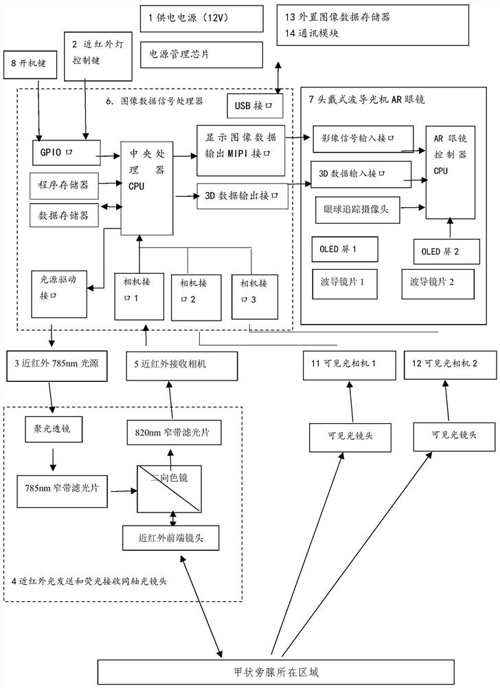
[0101] Embodiment 2 A position enhancement instrument constructed according to the identification enhancement method of the present invention, comprising:

[0102] The power supply 1 is used to supply power to the near-infrared autofluorescence excitation device and the data signal processing component, and its power supply port is electrically connected to the near-infrared autofluorescence excitation device and the data signal processing component respectively. A first switch 2 is connected, which is used to control the start and stop of the near-infrared autofluorescence excitation device; a second switch 8 is connected between the data signal processing component and the power supply, and is used to control the start and stop of the data signal processing component;

[0103] The near-infrared autofluorescence excitation device 3, its signal input end is connected to the signal of the data signal processing component, the power supply end is electrically connected to the pow...


PUM

PropertyMeasurementUnit
Thicknessaaaaaaaaaa

Abstract

A near-infrared fluorescence parathyroid gland recognition enhancement method based on an AR technology comprises the following steps of emitting near-infrared light of 785 nm to an area where parathyroid glands are located, generating autofluorescence of 820 nm by the parathyroid glands, and outputing position image coordinate information of the parathyroid glands in an image; synchronously acquiring left and right groups of image data acquired by a left camera and a right camera of a visible light RGB binocular camera, and solving the spatial positions of the left and right cameras and the actual spatial coordinates of each point in the visual field range; fitting 3D curved surfaces in view field ranges of a left image and a right image; projecting the position image coordinate information of the parathyroid gland onto a 3D curved surface in the visual field ranges, and solving actual space coordinates of the parathyroid gland; calculating an indication image to be projected on an AR display component in combination with the position of the AR display component; and achieving superposition overlapping of the indication image and the real scene through an observation window of the AR display component. The parathyroid gland recognition enhancement method has the advantages that the parathyroid gland position is rapidly detected and fused with an actual scene.

Description

technical field [0001] The invention relates to a method for identifying and enhancing near-infrared fluorescent parathyroid glands based on AR technology, which belongs to the field of medical operations. Background technique [0002] The parathyroid glands are small and changeable. In the visible light range, the parathyroid glands are similar to other tissues (such as fat) in color, and it is difficult to identify them during surgery. During thyroid surgery, the probability of parathyroid glands being injured or miscut is as high as 18-59% %. The protection of the parathyroid glands during the operation is extremely important to the recovery of the patient. Accidental injury can lead to hypoparathyroidism. The patient will suffer from numbness and convulsions due to calcium deficiency, which is extremely painful and requires calcium supplementation. The key to the protection of parathyroid glands is accurate identification. [0003] In addition, in some surgeries, the p...

Claims


Application Information

Patent Timeline
IPC IPC(8): A61B5/00A61B90/00
Inventor 俞志勇江兴智张乾威文仲杨青
Owner THE FIRST AFFILIATED HOSPITAL ZHEJIANG UNIV COLLEGE OF MEDICINE
Why Eureka
Try Eureka