Tissue pathology image cell nucleus segmentation method and system based on polar coordinate representation

A technology of histopathology and cell nucleus, applied in the field of image processing, can solve the problems of complex model structure, long training time, high hardware equipment requirements, etc., and achieve the effect of simple backbone network and low computational complexity

Pending Publication Date: 2021-02-05
SHANDONG NORMAL UNIV
View PDF8 Cites 2 Cited by
  • Summary
  • Abstract
  • Description
  • Claims
  • Application Information

AI Technical Summary

Problems solved by technology

However, the automatic segmentation of cell nuclei in pathological images usually has limitations such as low efficiency and poor results, and it is difficult to achieve fast instance segmentation of cell nuclei in pathological images.
Image target instance segmentation methods based on artificial intelligence, such as Mask-RCNN, can better solve the problem of cell nucleus segmentation in pathological images, but there are limitations such as complex model structure, long training time, and high hardware requirements.
[0004] In summary, there is still a lack of effective solutions to the problem of automatic detection of cell nuclei in histopathological images in existing technologies

Method used


Image

  • Tissue pathology image cell nucleus segmentation method and system based on polar coordinate representation
  • Tissue pathology image cell nucleus segmentation method and system based on polar coordinate representation
  • Tissue pathology image cell nucleus segmentation method and system based on polar coordinate representation

Examples


Embodiment 1

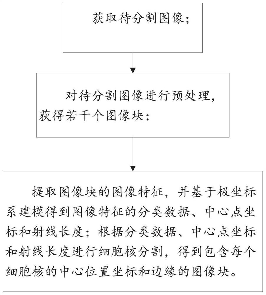
[0036] Such as figure 1 As shown, the disclosure provides a histopathological image nucleus segmentation method based on polar coordinate representation, including:

[0037] Obtain the image to be segmented;

[0038] Preprocessing the image to be segmented to obtain several image blocks;

[0039] Extract the image features of the image block, and obtain the classification data, center point coordinates, and ray length of the image features based on polar coordinate system modeling; perform cell nucleus segmentation according to the classification data, center point coordinates, and ray length, and obtain the center position containing each nucleus Coordinates and edges of the image patch.

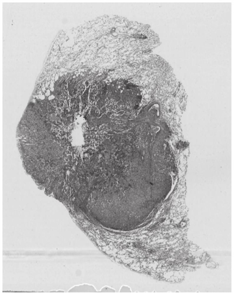
[0040] Further, the acquisition of the image to be segmented specifically includes obtaining pathological slices through paraffin fixation, sectioning, patching, H&E staining, and sealing, and then using a scanner to scan them into full-size digital pathological images (whole slide images...

Embodiment 2

[0058] A histopathological image nucleus segmentation system based on polar coordinate representation, including:

[0059] Data acquisition module: acquire the image to be segmented;

[0060] Image preprocessing module: preprocessing the image to be segmented to obtain several image blocks to be segmented;

[0061] Data processing module: extract the image features of the image block to be segmented, and obtain the classification data, center point coordinates, and ray length of the image feature based on polar coordinate system modeling; perform image block segmentation according to the classification data, center point coordinates, and ray length The nucleus is segmented to obtain an image block containing the coordinates of the center position and the edge of each nucleus.

[0062] Further, the specific manner in which the data acquisition module is configured corresponds to the specific steps of the method for segmenting cell nuclei in histopathological images based on po...

Embodiment 3

[0064] A computer-readable storage medium is used for storing computer instructions. When the computer instructions are executed by a processor, the method for segmenting cell nuclei in histopathological images based on polar coordinate representation as described in the above embodiments is completed.


PUM


Abstract

The invention provides a tissue pathology image cell nucleus segmentation method and system based on polar coordinate representation; wherein the method comprises the steps: extracting the image features of a to-be-segmented image block, and carrying out the modeling based on a polar coordinate system, and obtaining the classification data, central point coordinates and ray length of the image features; according to the classification data, the center point coordinates and the ray length, carrying out cell nucleus segmentation of the to-be-segmented image block to acquire the image block containing the center position coordinates and the edges of each cell nucleus. On the aspect of the processing effect, the invention provides the polar coordinate representation-based automatic cell nucleus segmentation method of the histopathological image for the first time. The method based on polar coordinates is very suitable for segmentation of circular objects; therefore, the pathological imagecell nucleus can be automatically segmented, the cell nucleus center and the ray length pointing to the target edge with the cell nucleus center as the starting point are obtained, and positioning andfine segmentation of the cell nucleus are achieved.

Description

technical field [0001] The present disclosure relates to the field of image processing, and in particular to a method and system for automatically segmenting cell nuclei based on histopathological images represented by polar coordinates. Background technique [0002] The precise positioning and segmentation of cell nuclei is an important step in pathological image analysis, which is used to quantitatively analyze the characteristics of tissues in digital pathological images, so as to realize computer-aided diagnosis. In recent years, artificial intelligence-based methods have shown great potential in pathological image analysis tasks. [0003] Many methods have been proposed for the segmentation of cells and nuclei in histopathological images, such as threshold-based image segmentation methods, edge-based image segmentation methods, and region-growing image segmentation methods. In addition, cell nucleus segmentation methods based on specific theories have also been proposed...

Claims


Application Information

Patent Timeline
Patent Type & Authority Applications(China)
IPC IPC(8): G06T7/12G06T7/13G16H30/20G06N3/04
CPCG06T7/12G06T7/13G16H30/20G06N3/045
Inventor 郑元杰马帅姜岩芸肖伟姚志刚周小明
Owner SHANDONG NORMAL UNIV
Why Eureka
Try Eureka