Application of MST1 as drug target in preparation of drug for treating colorectal cancer
A technology of colorectal cancer and drugs, applied in the field of biomedicine, can solve the problem of few clinical significance studies
- Summary
- Abstract
- Description
- Claims
- Application Information
AI Technical Summary
Problems solved by technology
Method used
Image
Examples
Embodiment 1
[0022] Example 1 Cell Culture and Transfection
[0023] 1 Materials and methods
[0024] 1.1 General information
[0025] A total of 81 colorectal cancer patients (tumor tissue and paired normal adjacent tissue) were collected from the Department of General Surgery of Nanchong Central Hospital from March 2009 to April 2014. This study has been approved by the Medical Ethics Committee and complies with the relevant requirements of the Declaration of Helsinki of the World Medical Association. All patients were aware of the research content and signed the informed consent.
[0026] 1.2 Cell culture and transfection
[0027] HCT116, SW480, LOVO and human colonic epithelial cells HCoEpiC were obtained from the Chinese Academy of Sciences (Shanghai) cell bank, and HCT116, SW480, LOVO cells were cultured in RPMI-1640 medium (Gibco, USA) with 10% fetal bovine serum. LV-MST1 and LV-NC lentiviral vectors were transfected according to the manufacturer's instructions (Gene Pharmaceuti...
Embodiment 2
[0049] The expression of embodiment 2MST1 in CRC tissue
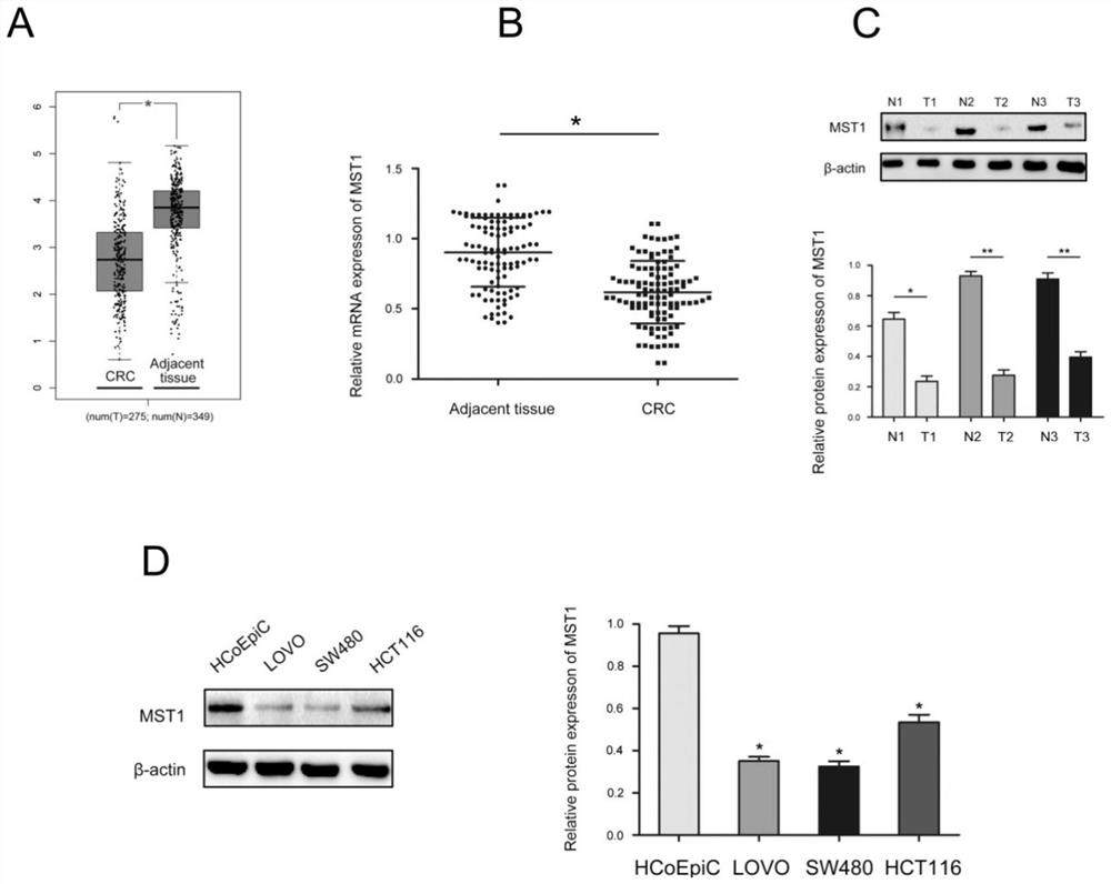
[0050] MST1 is lowly expressed in CRC tissues. First, 275 cases of CRC tissues and 349 cases of normal tissues were analyzed (http: / / gepia.cancer-pku.cn / index.html) to evaluate the level of MST1 in CRC. The results are shown in figure 1 .
[0051] figure 1 Middle A is the mRNA expression of MST1 in the TCGA database (colorectal cancer and adjacent non-cancerous tissues); B is the mRNA expression of MST1 in CRC and adjacent non-cancerous tissues; C is the protein expression of MST1 in adjacent cancerous tissues and adjacent non-cancerous tissues; D is the expression of MST1 in cells; *P<0.05, **P<0.01.
[0052] according to figure 1 The detection results showed that, compared with normal tissues, the expression of MST1 in CRC tissues was lower ( figure 1 A), qRT-PCR and Western blot method were used to detect MST1 expressing cells in CRC tissues. The mRNA level of MST1 in CRC tissues was significantly lower than tha...
Embodiment 3
[0053] Example 3 Correlation between immunohistochemical detection of MST1 and clinicopathological features of CRC
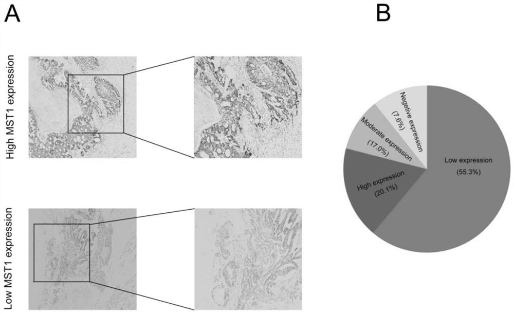
[0054] Immunohistochemical results such as figure 2 as shown, figure 2 Among them, A is the immunohistochemical analysis of MST1 expression in CRC and adjacent tissues; B is the statistical analysis of MST1 expression in CRC and adjacent tissues.
[0055] according to figure 2 The detection results showed that the low expression rate of MST1 in CRC was 61.1% (51 / 81) (Figure 2A and B). In addition, the positive expression of MST1 was mainly concentrated in the cytoplasm. By analyzing the relationship between MST1 and clinicopathological features of CRC patients, we found that low MST1 expression was positively correlated with tumor size (P=0.011) and differentiation (P<0.001).
PUM
 Abstract
Description
Claims
Application Information
 - R&D
- Intellectual Property
- Life Sciences
- Materials
- Tech Scout
- Unparalleled Data Quality
- Higher Quality Content
- 60% Fewer Hallucinations
Browse by: Latest US Patents, China's latest patents, Technical Efficacy Thesaurus, Application Domain, Technology Topic, Popular Technical Reports.
© 2025 PatSnap. All rights reserved.Legal|Privacy policy|Modern Slavery Act Transparency Statement|Sitemap|About US| Contact US: help@patsnap.com


