Eureka AIR delivers breakthrough ideas for toughest innovation challenges, trusted by R&D personnel around the world.

Liver cancer organoid and culture method, culture medium and application of organoid

A culture medium and organoid technology, applied in the field of biomedicine, can solve the problems of inability to simulate the penetration of drugs into cells, low accuracy, and different lethality

Active Publication Date: 2021-01-12
北京科途医学科技有限公司 +1
View PDF1 Cites 5 Cited by
  • Summary
  • Abstract
  • Description
  • Claims
  • Application Information

AI Technical Summary

Problems solved by technology

In addition, since the permeability of drugs to cells cannot be simulated by using two-dimensional monolayer cells, some drugs have different lethality to three-dimensional cultured cells and two-dimensional cultured monolayer cells.
[0004] Aiming at the problem that the accuracy of drug screening of existing liver cancer cell lines is not high and the primary liver cancer tissue cannot be subcultured in vitro, the development of a more authentic human liver cancer cell model can promote the precise drug use and drug screening of liver cancer. accuracy

Method used


Image

  • Liver cancer organoid and culture method, culture medium and application of organoid
  • Liver cancer organoid and culture method, culture medium and application of organoid
  • Liver cancer organoid and culture method, culture medium and application of organoid

Examples

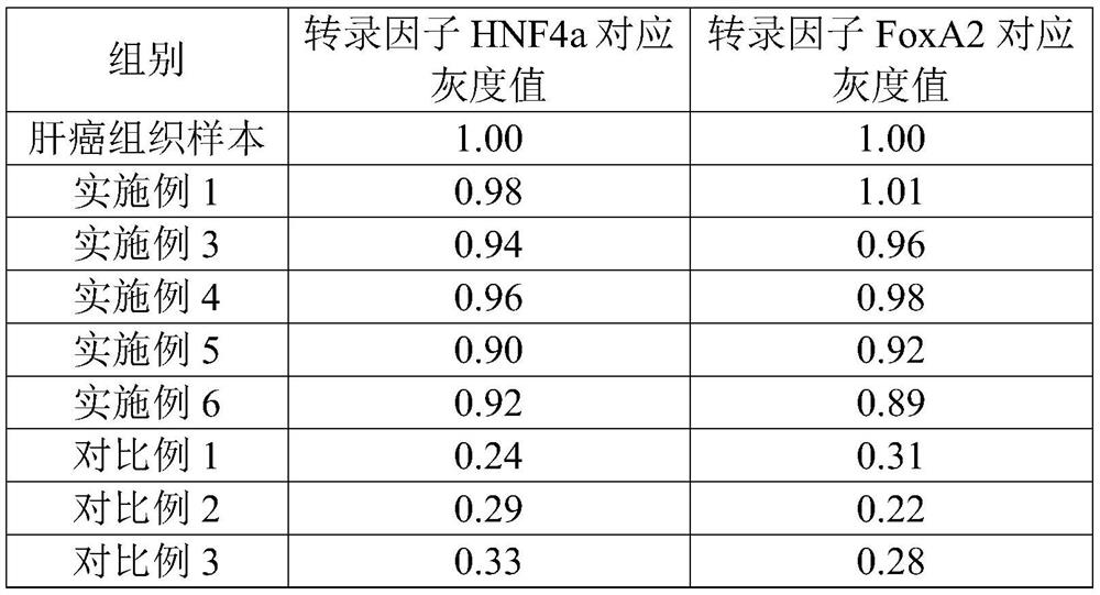

Embodiment 1

[0046] This example is used to illustrate the culture of human liver cancer organoids.

[0047] (1) Acquisition of human liver cancer tissue cells

[0048] Obtain cancer tissues from clinically diagnosed liver cancer patients as liver cancer tissue samples. Use pre-cooled PBS to clean the liver cancer tissue samples. After cleaning, use sterile instruments (such as surgical scissors) to remove the necrotic tissue in the liver cancer samples, and cut the liver cancer tissue samples into 1-2mm 3 Tissue blocks of different sizes were obtained to obtain liver cancer tissue blocks.

[0049] The obtained liver cancer tissue pieces were mixed with enzymatic hydrolysis solution (containing 10 mM HEPES, 100 μg / ml penicillin, 1x glutamine, 2 ng / ml collagenase and the rest of DMEM / F12 medium) to obtain the Enzymatic hydrolyzate, wherein 0.1 ml of enzymatic hydrolyzate is added for every 1 mg of liver cancer tissue block. Put the aforementioned digested product in an incubator at 37° C...

Embodiment 2

[0054] This example is used to illustrate the application of liver cancer organoids in drug screening.

[0055] The liver organoids cultured in Example 1 were digested with TrypLE Express for 1 min, and then DMEM high-glucose medium containing 2% by volume of FBS was added to stop the digestion, and the hydrolyzate was collected. The collected enzymatic solution was centrifuged at 300 g for 3 min, the supernatant was removed, the cell pellet was collected, and the number of cells after enzymatic hydrolysis was counted.

[0056] The above cell pellet was mixed with culture medium so that the concentration of liver cancer tissue cells was 6×10 3 Each cell is 50 μl, and then add 5% volume of Matrigel, and mix evenly on ice to obtain a culture to be cultured. Inoculate the culture to be cultured above into a 96-well low-adsorption plate, then place the 96-well plate in an incubator with 5% carbon dioxide at 37°C for 30 min, then add 40 μL of culture medium to each well of the 96-...

Embodiment 3

[0059]Liver cancer organoids were cultured according to the method of Example 1, except that the concentration of insulin growth factor-2 in the culture medium used in this example was 5 ng / ml.


PUM


Abstract

The invention relates to a method for culturing liver cancer organoid. The method comprises the following steps: mixing liver cancer tissue cells with a culture medium and matrix gel to obtain a to-be-cultured substance, wherein the culture medium containing an insulin growth factor-2, and the concentration of the insulin growth factor 2 being 1-50ng / ml, preferably 5-10ng / ml based on the culture medium; and carrying out culture amplification on the to-be-cultured substance to obtain the liver cancer organoid. According to the culture method of the liver cancer organoid provided by the invention, the adopted culture medium contains the insulin growth factor-2 with the specific concentration, and the insulin growth factor-2 with the specific concentration can promotes in-vitro growth of theliver cancer organoid, so that the culture success rate is relatively high when the method provided by the invention is used for culturing the liver cancer organoid.

Description

technical field [0001] The present disclosure relates to the technical field of biomedicine, in particular to a method for culturing liver cancer organoids, a medium for culturing liver cancer organoids, liver cancer organoids and their application in drug screening, precise drug detection and xenograft animal models Applications. Background technique [0002] The annual number of new cases of primary liver cancer in the world is as high as 854,000, of which the number of cases in China is 466,000, accounting for about 55% of the world. However, for the study of human liver cancer, there is still a lack of in vitro models that can truly reproduce the tumor pathophysiology of primary liver cancer. [0003] Common liver cancer cell lines have obvious differences in protein expression and xenograft tumorigenicity because they come from different individuals. In addition, since the liver cancer cell line was constructed with an immortalized gene, it is prone to dissimilation d...

Claims


Application Information

Patent Timeline
IPC IPC(8): C12N5/09C12Q1/02A01K67/027
CPCC12N5/0693C12N5/0671G01N33/5044A01K67/0271C12N2501/105C12N2500/60C12N2500/32C12N2501/119C12N2500/30C12N2503/02G01N2500/10A01K2207/12A01K2267/0331
Inventor 李程黄思颖孙志坚康平
Owner 北京科途医学科技有限公司
Why Eureka
Eureka Blog
Learn More