Pulmonary nodule detection method and device, model training method and device, equipment and medium
A detection method and technology of pulmonary nodules, applied in the field of image processing, can solve the problems of automatic detection missed detection, affecting detection results, false detection, etc.
- Summary
- Abstract
- Description
- Claims
- Application Information
AI Technical Summary
Problems solved by technology
Method used
Image
Examples
Embodiment 1
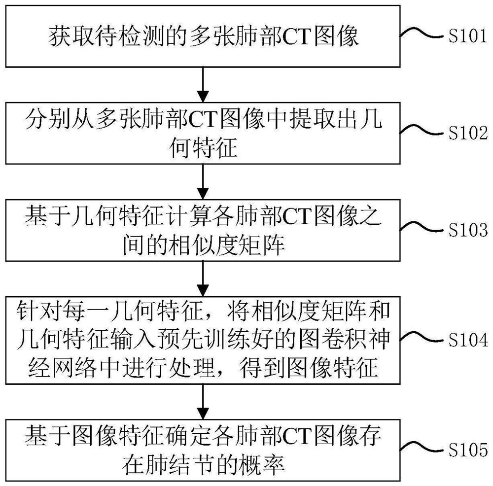
[0055] figure 1It is a flow chart of a pulmonary nodule detection method provided by Embodiment 1 of the present invention. This embodiment can be applied to the situation where pulmonary nodules in CT images are similar to blood vessels and other lung shadow structures, resulting in a large number of false positives. The method can be executed by the device for detecting pulmonary nodules provided by the embodiment of the present invention, which can be realized by software and / or hardware, and is usually configured in a computer device, such as figure 1 As shown, the method specifically includes the following steps:
[0056] S101. Acquire a plurality of lung CT images to be detected.
[0057] Specifically, CT (Computed Tomography) images, that is, computerized tomography images, use precisely collimated X-ray beams, γ-rays, ultrasonic waves, etc., to make a joint around a certain part of the human body together with a highly sensitive detector. A cross-sectional scan. The...
Embodiment 2
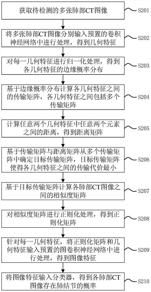
[0071] Figure 2A It is a flow chart of a method for detecting pulmonary nodules provided by Embodiment 2 of the present invention. This embodiment is refined on the basis of Embodiment 1 above, and describes in detail the process of extracting geometric features from lung CT images, The process of calculating the similarity matrix and the processing process of the graph convolutional neural network, such as Figure 2A As shown, the method includes:
[0072] S201. Acquire a plurality of lung CT images to be detected.
[0073] In a specific embodiment of the present invention, the lung CT image is a three-dimensional CT image with a size of 96×96×96, that is, the size of the lung CT image in three dimensions of length, width and height is 96 pixels. The three-dimensional CT image can make the original plane image become three-dimensional, and the density difference between the diseased tissue and the adjacent normal tissue is increased, and the condition of the diseased tissu...
Embodiment 3
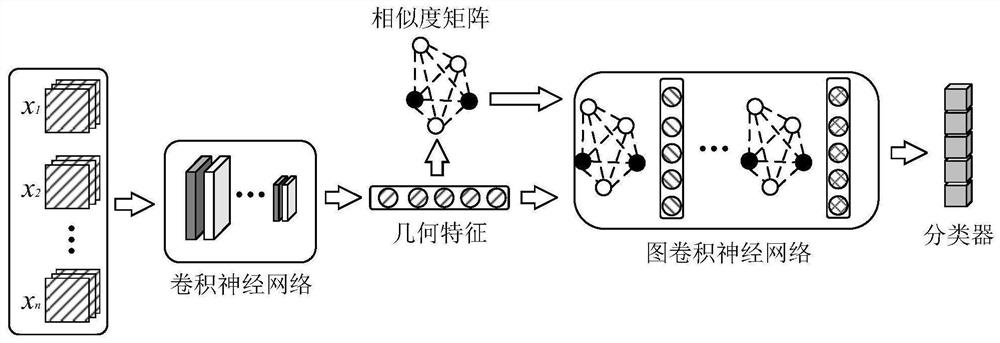
[0163] image 3 It is a flowchart of a pulmonary nodule detection model training method provided in Embodiment 3 of the present invention. This embodiment can be used for the training of the pulmonary nodule detection model provided in the embodiment of the present invention. The nodule detection model training device is implemented, and the device can be implemented by software and / or hardware, and is usually configured in a computer device. Such as image 3 As shown, the method specifically includes the following steps:
[0164] S301. Obtain a data set, where the data set includes a training set composed of multiple labeled lung CT image samples.
[0165] Specifically, in one of the embodiments of the present invention, the data set includes a training set composed of multiple labeled lung CT image samples, and the labels are used to indicate whether there are pulmonary nodules in the lung CT image samples. In the subsequent In the embodiment, we refer to the lung CT imag...
PUM
 Abstract
Description
Claims
Application Information
 - R&D Engineer
- R&D Manager
- IP Professional
- Industry Leading Data Capabilities
- Powerful AI technology
- Patent DNA Extraction
Browse by: Latest US Patents, China's latest patents, Technical Efficacy Thesaurus, Application Domain, Technology Topic, Popular Technical Reports.
© 2024 PatSnap. All rights reserved.Legal|Privacy policy|Modern Slavery Act Transparency Statement|Sitemap|About US| Contact US: help@patsnap.com


