Retinal vessel image segmentation method based on deep learning

A retinal blood vessel and image segmentation technology, applied in the field of image processing, can solve problems such as unsatisfactory segmentation methods, achieve good segmentation results, improve recognition accuracy, and improve feature utilization

Inactive Publication Date: 2020-10-30
DONGGUAN UNIV OF TECH
View PDF7 Cites 20 Cited by
  • Summary
  • Abstract
  • Description
  • Claims
  • Application Information

AI Technical Summary

Problems solved by technology

However, the advantages and disadvantages of the automatic segmentation method will directly lead to whether

Method used


Image

  • Retinal vessel image segmentation method based on deep learning
  • Retinal vessel image segmentation method based on deep learning
  • Retinal vessel image segmentation method based on deep learning

Examples


Embodiment Construction

[0050] The present invention will be described in further detail below through examples, and the following examples are explanations of the present invention and the present invention is not limited to the following examples.

[0051] like figure 1 Shown, a kind of retinal blood vessel image segmentation method based on deep learning of the present invention comprises the following steps:

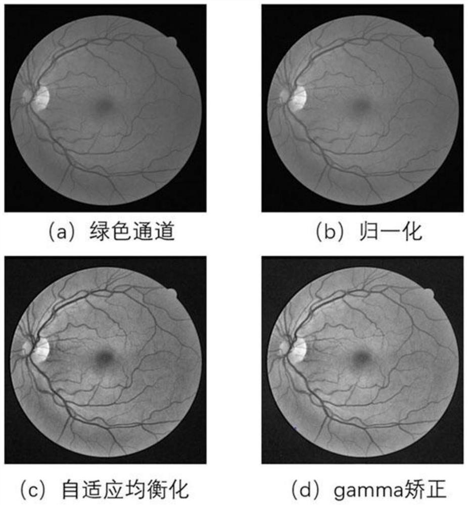
[0052] Step 1: fundus image enhancement, contrast enhancement is performed on the fundus image to highlight details of retinal blood vessels.

[0053] The processing of this part is mainly to improve the contrast between the retinal blood vessels and the background, making the blood vessels more prominent and improving the segmentation accuracy. Extract the green channel with higher contrast from the fundus image of the training set, and normalize it; then use adaptive histogram equalization, calculate the neighborhood histogram for each pixel in the image to obtain the histogram transform...


PUM


Abstract

The invention discloses a retinal vessel image segmentation method based on deep learning, and the method comprises the steps: carrying out the enhancement of a fundus image, amplifying the data of atraining set, constructing a dense connection convolution block, replacing a conventional convolution block with the dense connection convolution block, achieving the feature reuse, and improving thefeature extraction capability; constructing an attention mechanism module, and performing adaptive adjustment on the feature map to highlight important features so as to suppress invalid features; building a model, building a DA-Unet network, using the processed data set to perform training and parameter adjustment, and obtaining and storing an optimal segmentation model; and carrying out actual segmentation, segmenting the eye fundus image needing retinal vessel segmentation into 48 * 48 sub-block images by using a sliding window, inputting the 48 * 48 sub-block images into a DA-Uet network for segmentation, outputting segmented sub-block image results, and splicing the segmented small block images into a complete retinal vessel segmentation image. The blood vessel segmentation method canautomatically segment blood vessels and has a good segmentation effect on tiny blood vessels.

Description

technical field [0001] The invention relates to an image segmentation method, in particular to a deep learning-based retinal vessel image segmentation method, which belongs to the field of image processing. Background technique [0002] Changes in the morphology and structure of retinal blood vessels often indicate the emergence of certain pathological diseases, such as hypertension or diabetes. Hypertensive retinopathy is a retinal disease caused by high blood pressure, and its pathological features often manifest as increased curvature of retinal blood vessels or lead to vasoconstriction. Diabetic retinopathy is a retinal disease caused by elevated blood sugar, often accompanied by pathological features of retinal blood vessel swelling. Therefore, changes in retinal vascular structure in fundus images can assist ophthalmologists in discovering and diagnosing the early stages of some serious diseases. However, there are a large number of tiny blood vessels in the retinal ...

Claims


Application Information

Patent Timeline
IPC IPC(8): G06T7/00G06T7/10
CPCG06T7/0012G06T7/10G06T2207/30041G06T2207/30101G06T2207/20081G06T2207/20084G06T2207/20021
Inventor 赵晓芳陈雪芳林盛鑫梁桢灏
Owner DONGGUAN UNIV OF TECH
Why Eureka
Try Eureka