Breast cancer ultrasound image typing method and system fusing deep convolutional network and imaging omics features, and storage medium

A technology of ultrasound image typing and radiomics, applied in the field of ultrasound medical treatment, can solve the problems of low signal-to-noise ratio, low resolution and low accuracy, and achieve the effect of improving accuracy and accurate identification

Active Publication Date: 2020-08-25
HARBIN MEDICAL UNIVERSITY
View PDF18 Cites 8 Cited by
  • Summary
  • Abstract
  • Description
  • Claims
  • Application Information

AI Technical Summary

Problems solved by technology

However, due to the relatively low signal-to-noise ratio and resolution of ultrasound imaging, traditional feature extraction methods are difficult to obtain efficient expression of lesion features. Therefore, the accuracy of pathological classification of breast cancer using ultrasound images is relatively low. Therefore, a method is proposed. An accurate image processing, feature extraction and recognition method for ultrasound images of breast cancer to facilitate the use of ultrasound images by follow-up personnel is a technical problem that needs to be solved urgently in the market

Method used


Image

  • Breast cancer ultrasound image typing method and system fusing deep convolutional network and imaging omics features, and storage medium
  • Breast cancer ultrasound image typing method and system fusing deep convolutional network and imaging omics features, and storage medium
  • Breast cancer ultrasound image typing method and system fusing deep convolutional network and imaging omics features, and storage medium

Examples


Embodiment 1

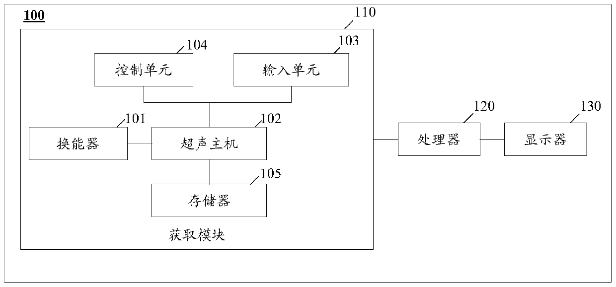
[0050] In an embodiment of the present invention, a system for classifying ultrasound images of breast cancer is provided, and the system includes an acquisition module 110 , a processor module 120 and a display module 130 . The acquiring module 110 acquires ultrasound data including the breast, the processor 120 processes the acquired ultrasound data, and analyzes to obtain the typing type of the ultrasound image of the breast, and the display 130 can display the acquired ultrasound data and the analyzed typing type.

[0051] Such as figure 1 As shown, the acquisition module 110 of this embodiment may be an ultrasound imaging device, that is, the ultrasound image or video is acquired by the ultrasound imaging device. Such as figure 1 As shown, the ultrasonic imaging device at least includes a transducer 101 , an ultrasonic host 102 , an input unit 103 , a control unit 104 and a memory 105 . The display screen of the ultrasound imaging device may be the display 130 of the sy...

Embodiment 2

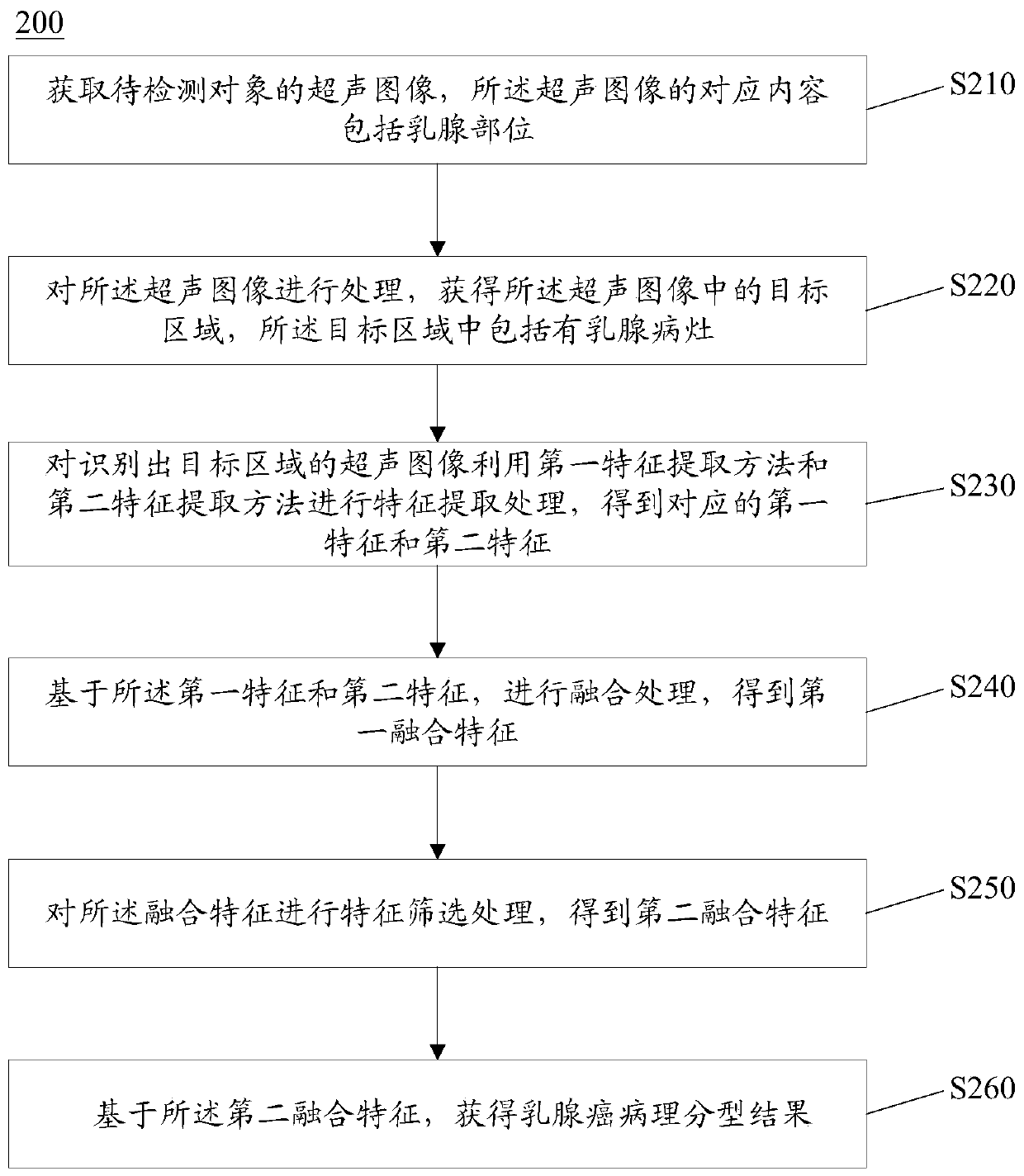
[0067] In one embodiment of the present invention, a classification method 200 for ultrasound images of breast cancer that combines deep convolutional networks and radiomics features is provided, which can be applied to ultrasound equipment, such as figure 2 As shown, the method 200 may include the following steps:

[0068] Step 210: Obtain an ultrasound image of the object to be detected, and the corresponding content of the ultrasound image includes breast parts.

[0069] In some embodiments, the ultrasound image of the object to be detected can be acquired through ultrasound equipment (such as color ultrasound equipment, black-and-white ultrasound equipment, etc.), database (such as PACS system), and the like.

[0070] Step 220: Process the ultrasonic image to obtain a target area in the ultrasonic image, and the target area includes breast lesions.

[0071] In some embodiments, the ultrasonic image may be processed by using a trained identification neural network model t...

Embodiment 3

[0132] In an embodiment of the present invention, a computer-readable storage medium is also provided, the computer-readable storage medium stores computer instructions, and the computer instructions are used to perform the fusion of the aforementioned deep convolutional network and radiomics of the present invention A characteristic sonographic classification method for breast cancer.

[0133] In addition, the implementation of the present invention can also be constructed in the form of a device, the device at least includes a processor and a storage device, the storage device stores instructions that can be read and executed by the processor, and the instructions use In order to realize and execute the ultrasonographic classification method of breast cancer by fusing deep convolutional network and radiomics features as described above.


PUM


Abstract

The invention provides a breast cancer ultrasound image typing method and system fusing a deep convolutional network and an imaging omics feature, and a computer readable storage medium. The method comprises the steps of acquiring an ultrasound image, wherein the corresponding content of the ultrasound image comprises a breast part; processing the ultrasonic image to obtain a target area in the ultrasonic image, the target area including a mammary gland lesion area image; extracting a first feature and a second feature from the ultrasonic image with the identified target area; performing fusion processing based on the first feature and the second feature to obtain a first fusion feature; performing feature screening processing on the first fusion feature to obtain a second fusion feature;and based on the second fusion feature, obtaining a typing result of the breast cancer ultrasonic image. According to the method, high-throughput ultrasonic image features and deep semantic features are extracted, fusion and feature screening are carried out, and effective and accurate recognition of the ultrasonic image is realized.

Description

technical field [0001] The invention relates to the field of ultrasonic medical technology, and belongs to the field of identification and processing of ultrasonic images, in particular to a breast cancer ultrasonic image identification and typing method and a corresponding system that integrates deep convolutional network and radiomics features. Background technique [0002] With the continuous development of medical equipment, ultrasonic imaging equipment has become one of the most widely used medical equipment and tools in clinical practice because of its non-invasive, real-time, convenient operation, and low price. The commonly used functional modes of ultrasound imaging include two-dimensional black and white (B) mode, spectral Doppler mode (PW / CW) and color flow mode (CF / PDI). The B mode relies on the amplitude of the ultrasonic echo signal for imaging, and what is obtained is the two-dimensional structure and shape information of the tissue. The greater the echo signa...

Claims


Application Information

Patent Timeline
Patent Type & Authority Applications(China)
IPC IPC(8): G06T7/40G06T7/13G06T7/00G06T5/50G06T5/20G06K9/46G06K9/62G06N3/04
CPCG06T7/0012G06T7/13G06T7/40G06T5/50G06T5/20G06T2207/10132G06T2207/20221G06T2207/30068G06T2207/30096G06V10/462G06N3/045G06F18/23G06F18/214
Inventor 田家玮张蕾王影俞卫东张云鹏时嘉欣
Owner HARBIN MEDICAL UNIVERSITY
Why Eureka
Try Eureka