Specific detection antigen of bovine echinococcosis granulosa and application of specific detection antigen
A technology of echinococcosis and antigen, which is applied in the field of medical devices, can solve problems such as cross-reaction, diagnostic error, and expensive preparation, and achieve the effects of strong specificity, rapid diagnosis, and high sensitivity
- Summary
- Abstract
- Description
- Claims
- Application Information
AI Technical Summary
Problems solved by technology
Method used
Image


Examples
Embodiment 1
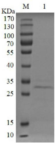
[0045] Embodiment 1, the preparation of Eg-H1 recombinant antigen
[0046] 1. Construction of pET-28a-Eg-H1 recombinant expression vector
[0047] In this embodiment, the nucleotide sequence of the Eg-H1 gene used is shown in SEQ ID NO.2, and the amino acid sequence of the protein is shown in SEQ ID NO.1. Eg-H1 is a multi-epitope recombinant antigen, and its epitopes are B-cell epitopes specific for two stages of Hexaconcus or Protoscoleia. Antigen B subunit 1, antigen B subunit 2, antigen Bsubunit 4, EG95, antigen protein, Ag5, EPC1, according to the bioinformatics analysis of their B cell epitope and secondary structure, intercepted some peptides and spliced them into recombinant proteins , which was named Eg-H1.
[0048] Construct the pET-28a-Eg-H1 expression vector, which was synthesized by Shanghai Biotechnology Co., Ltd., and the pET-28a-Eg-H1 expression vector was verified by double enzyme digestion and sequencing. The double enzyme digestion steps are as follows: ...
Embodiment 2
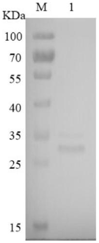
[0097] Example 2, Preparation of Hypothetical protein EGR_01530 recombinant antigen
[0098] 1. Construction of pET-28a-Hypothetical protein EGR_01530 expression vector
[0099] In this example, the nucleotide sequence of the Hypothetical protein EGR_01530 gene used is shown in SEQ ID NO.4, and the amino acid sequence of the protein is shown in SEQ ID NO.3. Hypothetical protein EGR_01530 antigen is a hypothetical protein containing a cysteine domain.
[0100] The prokaryotic expression vector pET-28a-Hypothetical protein EGR_01530 expression plasmid was constructed. The expression vector was synthesized by Shanghai Biotechnology Co., Ltd. The double enzyme digestion steps are as follows:
[0101] (1) Centrifuge the centrifuge tube containing the lyophilized powder of the expression vector of the target gene at 3000 rpm / normal temperature for 1 min.
[0102] (2) with 50 μL sterile ddH 2 O Dissolve the lyophilized powder, then mix gently with a vortex instrument, and centri...
Embodiment 3
[0109] Example 3, Preparation of bovine echinococcosis time-resolved fluorescent immunochromatographic test strips
[0110] Such as Figure 7 As shown, the time-resolved fluorescent immunochromatography test strip provided in this embodiment comprises a PVC base plate and a sample pad 2 positioned on the PVC base plate 1, a binding pad 3, a nitrocellulose film 5 (NC membrane, a chromatographic membrane), an absorbent paper 4. One end of the nitrocellulose membrane is connected to one end of the binding pad, the other end of the nitrocellulose membrane is connected to one end of the absorbent paper, one end of the sample pad is connected to the other end of the binding pad; the binding pad is coated with time-resolved fluorescently labeled Eg - For H1 recombinant antigen, a detection line 6 is set on the side of the nitrocellulose membrane close to the binding pad, a quality control line 7 is set on the layer close to the absorbent pad, and the detection line 6 is coated with ...
PUM
 Abstract
Description
Claims
Application Information
 - R&D
- Intellectual Property
- Life Sciences
- Materials
- Tech Scout
- Unparalleled Data Quality
- Higher Quality Content
- 60% Fewer Hallucinations
Browse by: Latest US Patents, China's latest patents, Technical Efficacy Thesaurus, Application Domain, Technology Topic, Popular Technical Reports.
© 2025 PatSnap. All rights reserved.Legal|Privacy policy|Modern Slavery Act Transparency Statement|Sitemap|About US| Contact US: help@patsnap.com