
System for rapidly detecting new coronavirus 2019-nCoV in blood sample and preparation method of system

A 2019-ncov, coronavirus technology, applied in the fields of medical testing and immunodiagnosis systems, can solve the problems of complex operation and difficult large-scale screening, and achieve simple operation, reduce non-specific adsorption, and reduce interference background. Effect

Pending Publication Date: 2020-06-12
北京大弘生物技术有限公司
View PDF4 Cites 18 Cited by
  • Summary
  • Abstract
  • Description
  • Claims
  • Application Information

AI Technical Summary

Problems solved by technology

[0006] Recently, researchers across the country have done a lot of research work on the detection of new coronavirus (2019-nCoV) infection, and quickly have a number of fluorescent quantitative PCR detection reagents and emergency listings. However, PCR fluorescent quantitative detection kits have operational Complex and other characteristics, it is difficult to use for large-scale screening

Method used


Image

  • System for rapidly detecting new coronavirus 2019-nCoV in blood sample and preparation method of system
  • System for rapidly detecting new coronavirus 2019-nCoV in blood sample and preparation method of system

Examples


Embodiment 1

[0032] Embodiment 1 Rapid novel coronavirus IgM and IgG joint detection system of the present invention and its preparation

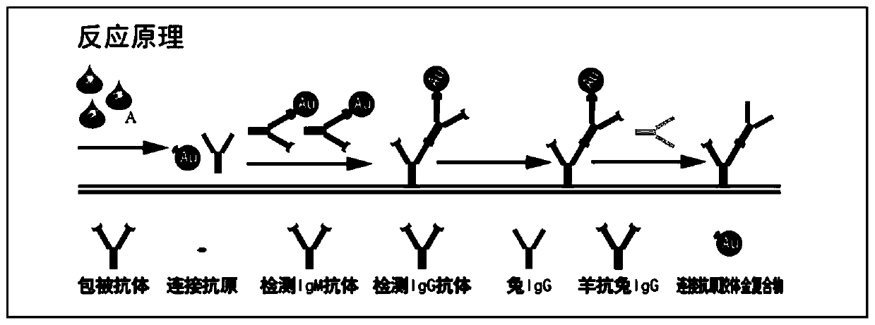
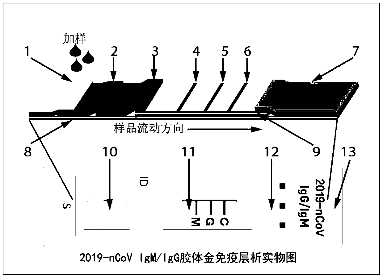
[0033] Such as Figure 1-2 As shown, the rapid new coronavirus IgM and IgG combined detection system of the present invention includes a buckle 13, a colloidal gold immunochromatography test strip 12 and a sample buffer, wherein the buckle 13 is a colloidal gold immunochromatography test strip The outer shell structure includes a sample injection hole 10 and an observation window 11 .

[0034] The structure of the colloidal gold immunochromatographic test strip is as follows: figure 2 As shown, it includes sample pad 1; marker pad 3; absorbent pad 7; PVC backing sheet 8; and NC membrane 9. When testing a whole blood sample, the test strip also includes a whole blood separation membrane 2 . Among them, the sample pad 1 is placed on the whole blood separation membrane 2, the whole blood separation membrane 2 is placed on the marking pad 3, the marking...

Embodiment 2

[0044] Example 2: Rapid detection of new coronavirus IgM and IgG in blood samples by the rapid new coronavirus IgM and IgG joint detection system of the present invention

[0045] 1. Colloidal gold and new coronavirus N protein link antigen labeling

[0046] Take 10ml of colloidal gold, use 0.2M NaHCO3 buffer pH8.0, add 100μg of new coronavirus N protein-linked antigen, mix well, let stand for 20min, add 5% BSA 1ml and mix well, centrifuge at 4℃, 10000r / min for 1h, discard Clear, dissolve the precipitate with TBS buffer to 10ml, centrifuge at 4°C, 10,000r / min for 1h, discard the supernatant, dilute the precipitate to 1ml with TBS, and obtain colloidal gold-labeled new coronavirus N protein-linked antigen;

[0047] The same procedure was used to prepare quality control molecules, colloidal gold-labeled rabbit IgG.

[0048] 2. System construction

[0049] a) Marker pad preparation

[0050] Mix the two colloidal gold labeling complexes at a ratio of 1:1, and add bovine serum a...


PUM


Abstract

The invention provides a system for rapidly detecting new coronavirus 2019-nCoV in a blood sample. The system is used for detecting IgM and IgG antibodies of the new coronavirus 2019-nCoV, and comprises a buckle, a colloidal gold immunochromatography test strip and a sample buffer solution. The colloidal gold immunochromatography-free test strip comprises a sample pad, a whole blood separation membrane, a marking pad, a chromatography membrane, a water absorption pad and a bottom plate which are connected in sequence, a colloidal gold-new coronavirus N protein connecting antigen compound and acolloidal gold-quality control molecular compound are fixed on the marking pad at the same time, and a mouse anti-human IgM-mu chain antibody and a mouse anti-human IgG-mu chain antibody are respectively fixed on a detection line. During detection, after the blood sample to be detected is added to the sample pad, the sample buffer solution is added; if the sample buffer solution is not used, butthe sample is directly detected, the new coronavirus IgM and IgG in the sample cannot be detected or false positive easily occurs, so that the existence of the sample buffer solution is crucial for realizing the detection of the sample.

Description

technical field [0001] The invention relates to the field of medical testing and immunodiagnosis systems, in particular to a system for rapidly detecting the new coronavirus 2019-nCoV in blood samples. Background technique [0002] The coronavirus was first isolated from chickens in 1937. In 1965, Tyrrell and Bynoe used the ciliated trachea tissue of the embryo to cultivate the coronavirus for the first time. This virus can be seen under the electron microscope as a corona-like peripheral crown, so it is called For coronavirus (Coronaviridae). The diameter of coronavirus particles is 60-200nm, with an average diameter of 100nm, spherical or oval, and pleomorphic. Viruses of the genus Coronavirus are positive-sense single-stranded RNA viruses with an envelope. The genetic material is all RNA. There are spines on the envelope. The entire virus is like a corona. The spines of different coronaviruses are significantly different. Among them, a variant of the coronavirus is the ...

Claims


Application Information

Patent Timeline
IPC IPC(8): G01N33/569G01N33/558
CPCG01N33/56983G01N33/558
Inventor 潘文东李道峰
Owner 北京大弘生物技术有限公司
Learn More