
Heart failure animal model for implanting pacemaker through external jugular vein under guidance of B-mode ultrasound

An animal model and pacemaker technology, applied in veterinary surgery, veterinary instruments, cardiac stimulators, etc., can solve the problems of single model function, difficult to determine the implantation position of pacemaker electrodes, complex structure, etc. into precise effects

Inactive Publication Date: 2020-05-29
THE FIRST TEACHING HOSPITAL OF XINJIANG MEDICAL UNIVERCITY
View PDF3 Cites 0 Cited by
  • Summary
  • Abstract
  • Description
  • Claims
  • Application Information

AI Technical Summary

Problems solved by technology

[0003] Due to the complex internal structure of the heart, it is difficult to determine the electrode implantation position of the pacemaker, which requires the use of a model to assist the experiment. The existing model has a single function and cannot assist in the implantation of the pacemaker, so it is difficult to determine the implantation position. , the present invention proposes an animal model of heart failure in which a pacemaker is implanted through the external jugular vein under the guidance of B-ultrasound to solve the problems existing in the prior art

Method used


Image

  • Heart failure animal model for implanting pacemaker through external jugular vein under guidance of B-mode ultrasound
  • Heart failure animal model for implanting pacemaker through external jugular vein under guidance of B-mode ultrasound
  • Heart failure animal model for implanting pacemaker through external jugular vein under guidance of B-mode ultrasound

Examples


Embodiment Construction

[0020] In order to deepen the understanding of the present invention, the present invention will be further described below in conjunction with the examples, which are only used to explain the present invention, and do not constitute a limitation to the protection scope of the present invention.

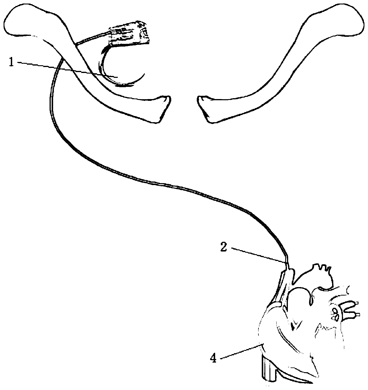
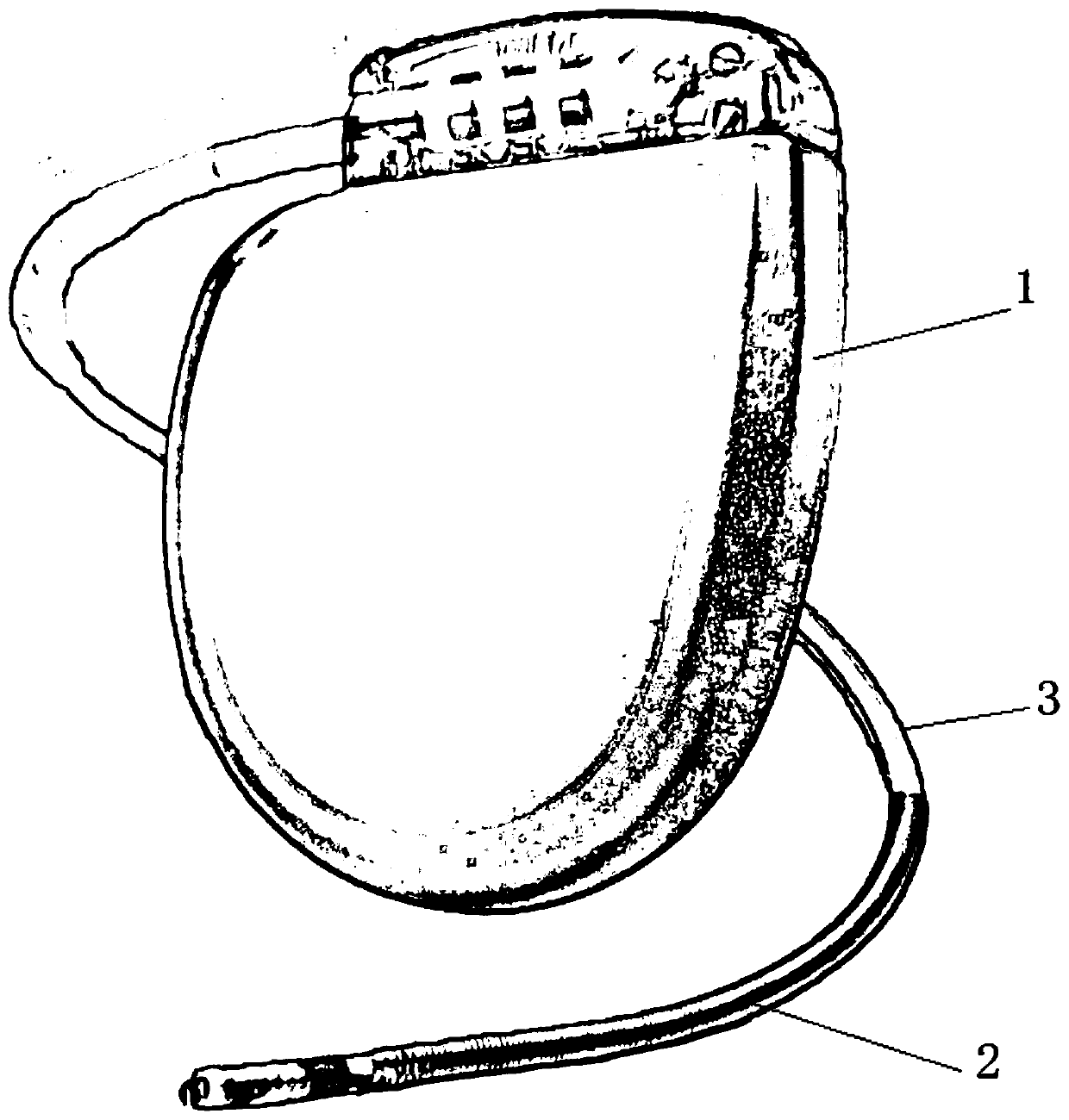
[0021] according to figure 1 , 2 , 3, the present embodiment provides an animal model of heart failure in which a pacemaker is implanted through the external jugular vein under the guidance of B-ultrasound, including an animal model of heart failure, a pacemaker 1, a spiral electrode 2, and a peelable sheath 3. Electrophysiological recorder and cardiac ultrasound instrument, the spiral electrode is inserted into the strippable sheath, and the strippable sheath pulls the spiral electrode, the heart failure animal model is provided with a heart 4, the The inner end of the helical electrode is located at the site of implantation in the right ventricle of the heart.

[0022] The outer ...


PUM


Abstract

The invention discloses a heart failure animal model for implanting a pacemaker through an external jugular vein under the guidance of B-mode ultrasound. The heart failure animal model comprises a heart failure animal model, the pacemaker, a spiral electrode, a strippable sheathing canal, an electrophysiological recorder and a cardiac ultrasound instrument, wherein the spiral electrode is insertedinto the strippable sheathing canal which pulls the spiral electrode; a heart is arranged in the heart failure animal model; and the inner end of the spiral electrode is located at a right ventricleimplantation position of the heart. According to the invention, the cardiac ultrasonic instrument is used for positioning; the electrophysiological recorder is used for sending stimulation; the innerend of the spiral electrode is implanted into the right ventricle of the heart according to the voltage of the electrophysiological recorder; the implantation position of the spiral electrode is determined when the waveform of a pacing electrocardiogram displayed by the cardiac ultrasonic instrument is a wide deformity QRS wave; meanwhile, when the heart rate and the pacing frequency displayed bythe electrophysiological recorder are consistent, the spiral electrode is fixed on the right ventricular muscle of the heart, so the implantation position is convenient to be determined.

Description

technical field [0001] The invention relates to the technical field of models, in particular to an animal model of heart failure in which a pacemaker is implanted through an external jugular vein under the guidance of B-ultrasound. Background technique [0002] In some medical experiments, it is often necessary to use animal models to do some experiments. Among them, in the field of heart, a pacemaker is needed for heart failure, and the implantation of a pacemaker is a major problem; [0003] Due to the complex internal structure of the heart, it is difficult to determine the electrode implantation position of the pacemaker, which requires the use of a model to assist the experiment. The existing model has a single function and cannot assist in the implantation of the pacemaker, so it is difficult to determine the implantation position. The present invention proposes an animal model of heart failure in which a pacemaker is implanted through the external jugular vein under t...

Claims


Application Information

Patent Timeline
IPC IPC(8): A61D1/00A61N1/362A61N1/365A61N1/37
CPCA61D1/00A61N1/362A61N1/36585A61N1/36592A61N1/3702
Inventor 汤宝鹏张玲周贤惠芦颜美李耀东张疆华邢强祖克拉·吐尔洪
Owner THE FIRST TEACHING HOSPITAL OF XINJIANG MEDICAL UNIVERCITY
Learn More