
A method for automatically identifying radiotherapy-at-risk organs in CT images based on a deep semantic network

A semantic network and CT image technology, applied in the field of medical image processing, can solve the problems of low delineation efficiency, inconsistent delineation results, and poor repeatability, and achieve the effect of improving generalization performance and work efficiency.

Active Publication Date: 2021-06-04
PERCEPTION VISION MEDICAL TECH CO LTD
View PDF9 Cites 0 Cited by
  • Summary
  • Abstract
  • Description
  • Claims
  • Application Information

AI Technical Summary

Problems solved by technology

Manual drawing by doctors has the following disadvantages: 1. The drawing efficiency is low; 2. It relies heavily on the doctor's clinical experience; 3. The repeatability is poor, and the results drawn by different doctors at different times and under different conditions are inconsistent.

Method used


Image

  • A method for automatically identifying radiotherapy-at-risk organs in CT images based on a deep semantic network
  • A method for automatically identifying radiotherapy-at-risk organs in CT images based on a deep semantic network
  • A method for automatically identifying radiotherapy-at-risk organs in CT images based on a deep semantic network

Examples


Embodiment Construction

[0062] In order to make the purpose, technical solutions and advantages of the embodiments of the present invention clearer, the technical solutions in the embodiments of the present invention will be clearly and completely described below in conjunction with the drawings in the embodiments of the present invention. Obviously, the described embodiments It is a part of embodiments of the present invention, but not all embodiments. Based on the embodiments of the present invention, all other embodiments obtained by persons of ordinary skill in the art without creative efforts fall within the protection scope of the present invention.

[0063] Unless expressly stated otherwise, throughout the specification and claims, the term "comprise" or variations thereof such as "includes" or "includes" and the like will be understood to include the stated elements or constituents, and not Other elements or other components are not excluded.

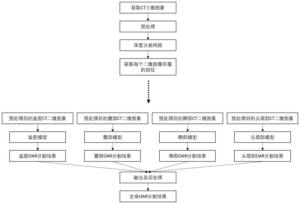
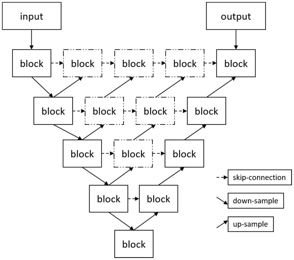
[0064] figure 1 A flowchart of a method for au...


PUM


Abstract

An embodiment of the present invention provides a method for automatically identifying organs at risk for radiotherapy in CT images based on a deep semantic network. The method includes the following steps: Step S1: Preprocessing the CT three-dimensional image; Step S2: Obtaining each The position to which two two-dimensional images belong; Step S3: Construct the deep semantic segmentation models for the pelvic cavity, abdomen, chest, and head and neck respectively; Step S4: Input the two-dimensional images belonging to the pelvic cavity, abdomen, chest, and head and neck respectively A trained deep semantic segmentation model for the corresponding pelvic cavity, abdomen, chest, and head and neck is used to identify the organs at risk for radiotherapy; step S5: the output results of the deep semantic segmentation models for the pelvic cavity, abdomen, chest, and head and neck are performed merge. This method implements artificial intelligence-assisted outline delineation of organs at risk in radiotherapy in the workflow of radiotherapy planning, preoperative evaluation and surgical planning, which can effectively improve the work efficiency of medical workers.

Description

technical field [0001] The invention relates to the technical field of medical image processing, in particular to a method for automatically identifying organs at risk for radiotherapy in CT images based on a deep semantic network. Background technique [0002] In the field of medicine, precision radiation therapy technology has greatly improved the survival rate of cancer patients. However, these advanced treatment methods require not only accurate judgment of the contour of the target tumor, but also accurate recognition of the contours of vital organs around the tumor, so that these organs can be protected during radiation therapy. In addition, in the field of surgical applications, accurate preoperative assessment and standardized radical surgery are important measures to improve the efficacy of tumor diagnosis and treatment. Organ contour recognition based on CT image data can help doctors complete surgical planning quickly, accurately and with high consistency. Steps ...

Claims


Application Information

Patent Timeline
Patent Type & Authority Patents(China)
IPC IPC(8): G06T7/00G06T7/11G06T7/136G06K9/46G06K9/62
CPCG06T7/0012G06T7/11G06T7/136G06T2207/10081G06T2207/20081G06T2207/20084G06T2207/30004G06V10/464G06F18/24
Inventor 魏军朱德明李松峰陈昌秀蒋雪田孟秋
Owner PERCEPTION VISION MEDICAL TECH CO LTD
Learn More