
Imaging probe, and preparation method and application thereof

An imaging probe and self-assembly technology, applied in the fields of tumor imaging and medicine, can solve problems such as limiting the application of intraoperative navigation technology, achieve long-term retention effects, increase fluorescence signal-to-noise ratio, and break through low fluorescence signal-to-noise ratio Effect

Inactive Publication Date: 2020-01-10
THE NAT CENT FOR NANOSCI & TECH NCNST OF CHINA
View PDF5 Cites 7 Cited by
  • Summary
  • Abstract
  • Description
  • Claims
  • Application Information

AI Technical Summary

Problems solved by technology

However, almost all fluorescent molecules approved by the Food and Drug Administration (FDA), such as indocyanine green (ICG), 5-aminolevulinic acid (5-ALA), and methylene blue (MB), etc., are Metabolic features and high signal background in organs with excretory function such as the kidney, which greatly limits the application of intraoperative navigation technology in renal cancer surgery

Method used


Image

  • Imaging probe, and preparation method and application thereof
  • Imaging probe, and preparation method and application thereof
  • Imaging probe, and preparation method and application thereof

Examples


Embodiment 1

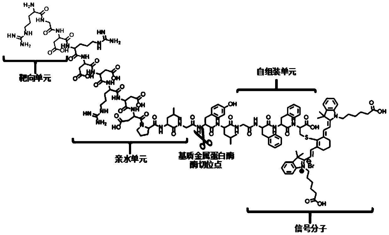
[0063] This embodiment provides an imaging probe RGDRDDRDDPGYLGFFC-Cy, whose structural formula can be represented by formula I, and the probe is prepared by polypeptide solid-phase synthesis.

[0064] Utilize the step that described probe is synthesized by polypeptide solid-phase synthesis method as follows:

[0065] (1) Synthesis of the polypeptide sequence RGDRDDRDDPGYLGFFC (hereinafter referred to as "polypeptide 1")

[0066] Experimental instruments and materials:

[0067] Dimethylformamide (DMF), piperidine, Wang resin, dichloromethane (DCM), ninhydrin reagents (ninhydrin, vitamin C, and phenol), benzotriazole-N,N,N' ,N'-tetramethyluronium hexafluorophosphate (HBTU), hexahydropyridine, triisopropylsilane (TIS), ethanedithiol (EDT), anhydrous ether, trifluoroacetic acid (TFA), N- Methylmorpholine (NMM), N-fluorenylmethoxycarbonyl-6-aminocaproic acid (Fmoc-e-Acp-OH), methanol, Fmoc-cysteine ​​(Fmoc-Cys(Trt)-OH), Fmoc -Aspartic acid (Fmoc-Asp(OtBu)-OH), Fmoc-phenylalanin...

Embodiment 2

[0084] This embodiment provides an imaging probe RGDPGYLGFFC-Cy, which can be represented by formula II, and the preparation method of the probe is the same as that in Example 1.

[0085] The polypeptide sequence RGD is the target recognition unit, PLGYLG is the enzyme hydrolysis substrate unit, the enzyme hydrolysis site is located between G and Y, FFC is the self-assembly unit, and C is used to connect the fluorescent group Cy.

Embodiment 3

[0087] This embodiment provides an imaging probe SCYNTNHVPLSPKYGPAKLVFFC-Cy, which can be represented by formula III, and its preparation method is the same as that of Example 1.

[0088] The receptor corresponding to the fragment SCYNTNHVPLSPKY in the polypeptide is carbonic anhydrase-9, the fragment GPA is the hydrolysis substrate unit of fibroblast activation protein-α, the molecular cleavage site is located between P and A, and the polypeptide sequence KLVFFC is a self-assembled fragment. Amino acid C is used to attach the fluorophore.


PUM


Abstract

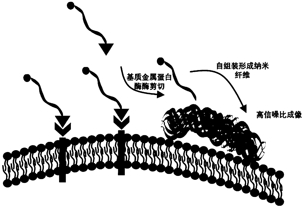
The invention provides an imaging probe, which comprises a targeted identification unit, an enzyme hydrolysis substrate unit, a self-assembling unit and a signal molecule, wherein the enzyme hydrolysis substrate unit is a peptide sequence which contains a functional enzyme substrate, the peptide sequence is PLGYLG or GPA, the targeted identification unit, the enzyme hydrolysis substrate unit and the self-assembling unit are connected in sequence through amido bonds, and the signal molecule is connected with the self-assembling unit. The imaging probe provided by the invention has a tumor targeting function and an assembling function, meanwhile, the imaging probe can generate efficient enzyme digestion reaction with high-expression enzymes in a tumor micro-environment on a tumor focus position through the enzyme hydrolysis substrate unit, so that self-assembling is carried out to form a specific nanometer fiber, long-acting retention is realized, the signal-to-noise ratio of tumors on organs, including a kidney, a liver, a bladder and the like, can be obviously improved, and a new method is provided for the intraoperative navigation excision of organ tumors.

Description

technical field [0001] The invention belongs to the technical field of medicine, specifically relates to the field of tumor imaging, in particular to an imaging probe and its preparation method and application. Background technique [0002] Because renal cell carcinoma is not sensitive to chemotherapy and radiotherapy, surgery has become the most effective treatment for renal cell carcinoma. Currently, radical nephrectomy (RN) and nephron-sparing partial nephrectomy (NSS) are the two main surgical approaches for the treatment of RCC. Compared with RN, NSS has been widely proven to significantly preserve renal function in patients while improving patient survival. However, the positive surgical margin rate (PSM) after NSS is high, and there are difficult-to-detect lesions in some complex lesions, which often lead to recurrence after surgery. Therefore, how to achieve precise tumor resection during NSS is a key issue for surgeons. [0003] Traditional imaging methods such a...

Claims


Application Information

Patent Timeline
Patent Type & Authority Applications(China)
IPC IPC(8): A61K49/00C07K7/08C07K7/02C07K1/04
CPCA61K49/0056A61K49/0069C07K7/02C07K7/08
Inventor 王浩徐万海安红维侯大勇王子琦王佳起赵昌浩
Owner THE NAT CENT FOR NANOSCI & TECH NCNST OF CHINA
Learn More