
Method and device for determination of imaging modality and parameters thereof, and imaging system

An imaging system and imaging technology, applied in image data processing, neural learning methods, medical images, etc., can solve the problems of infeasible diagnosis, limited diagnosis, re-shooting, etc.

Active Publication Date: 2019-11-26
SIEMENS HEALTHCARE GMBH
View PDF4 Cites 5 Cited by
  • Summary
  • Abstract
  • Description
  • Claims
  • Application Information

AI Technical Summary

Problems solved by technology

Especially in the most frequently used sequences in clinical practice, fast spin echo sequences, ghosting artifacts frequently occur in the event of patient motion, which limit the diagnosis, make it completely impossible or require re-imaging

Method used


Image

  • Method and device for determination of imaging modality and parameters thereof, and imaging system
  • Method and device for determination of imaging modality and parameters thereof, and imaging system

Examples


Embodiment Construction

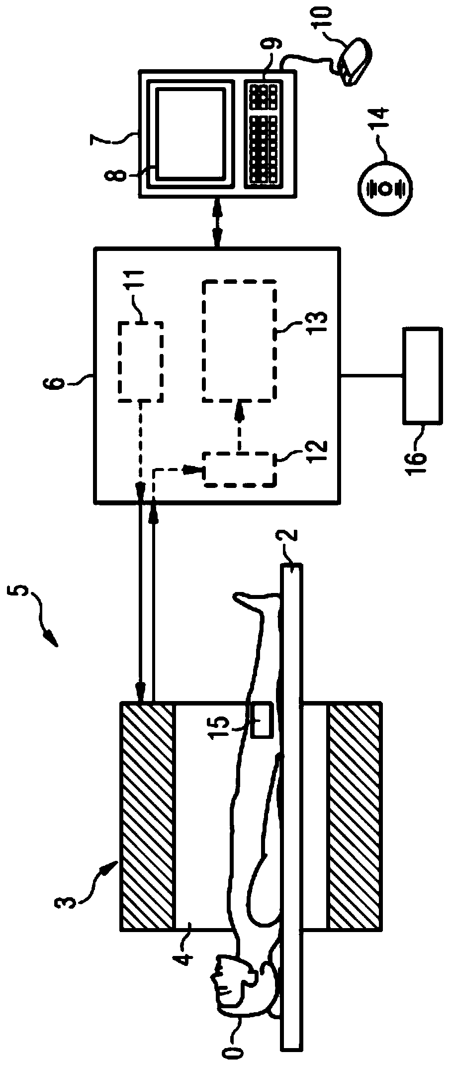
[0089] exist figure 1 The imaging system according to the present invention is shown in, figure 1 A device 16 according to the invention for determining the medical imaging modality and the parameters to be used here for the determined imaging modality is shown schematically next to the magnetic resonance system 5 (as a medical imaging modality). The magnetic resonance system 5 basically comprises: a tomograph 3 , by means of which the magnetic field required for the magnetic resonance examination is generated in the measurement chamber 4 ; a table or bed 2 ; a control device 6 , by means of which The tomography scanner 3 is controlled and the magnetic resonance data is detected by the tomography scanner 3 ; and the terminal 7 connected to the control device 6 .

[0090] The control device 6 for its part comprises an actuation unit 11 , a receiving device 12 and an evaluation device 13 . During the creation of the image data record, magnetic resonance data are detected by th...


PUM


Abstract

The invention relates to a medical imaging modality and a determination of parameters to be used for the determined imaging modality in order to create an image of an examination object (O) by means of the determined imaging modality and the determined parameters. Here, the following steps are carried out: information obtained from a pre-examination of the examination object (O) is automatically classified in order to obtain a classification result relating to an interference effect that can be expected when the image is created; and the classification result is analyzed so as to evaluate theresult. The imaging modality and the parameter are determined in dependence on the result of the evaluation, such that a classified interference influence is minimized when an image is generated fromthe examination object (O) by means of the determined imaging modality and the determined parameter.

Description

technical field [0001] The invention relates to the determination of an imaging modality and the determination of parameters of the determined imaging modality. Background technique [0002] Medical imaging modalities, such as magnetic resonance tomography, computed tomography, positron emission tomography, single photon emission computed tomography or radiographs, are susceptible to patient-specific disturbances in different ways and methods. The most disturbing influence here is patient movement, which in all imaging modalities leads to motion artifacts in the generated images. In computed tomography, such patient movements appear, for example, as streaks or ghosts, whereas in magnetic resonance imaging patient movements can act in different ways depending on the imaging frequency and the set parameters. Especially in the most frequently used sequences in clinical practice, the fast spin echo sequences, ghosting artifacts frequently occur during patient movement, which li...

Claims


Application Information

Patent Timeline
Patent Type & Authority Applications(China)
IPC IPC(8): G06T11/00G06N3/08A61B5/055A61B6/00A61B6/03A61B8/00
CPCG06T11/008G06N3/08A61B5/055A61B6/00A61B6/032A61B6/037A61B8/00G06T2207/10088G06T2207/10101A61B8/5215A61B6/5211A61B5/113A61B5/7264A61B5/7221G16H30/40G16H20/40A61B5/318A61B5/7292A61B5/7207A61B8/13A61B5/746A61B5/024
Inventor 马里奥·策勒
Owner SIEMENS HEALTHCARE GMBH
Learn More