SD-OCT image macular fovea centralis center positioning method
A center positioning and center position technology, applied in image enhancement, image analysis, image data processing, etc., can solve problems such as failure and poor retinal robustness, and achieve the effect of improving accuracy and reducing labor.
- Summary
- Abstract
- Description
- Claims
- Application Information
AI Technical Summary
Problems solved by technology
Method used
Image
Examples
Embodiment
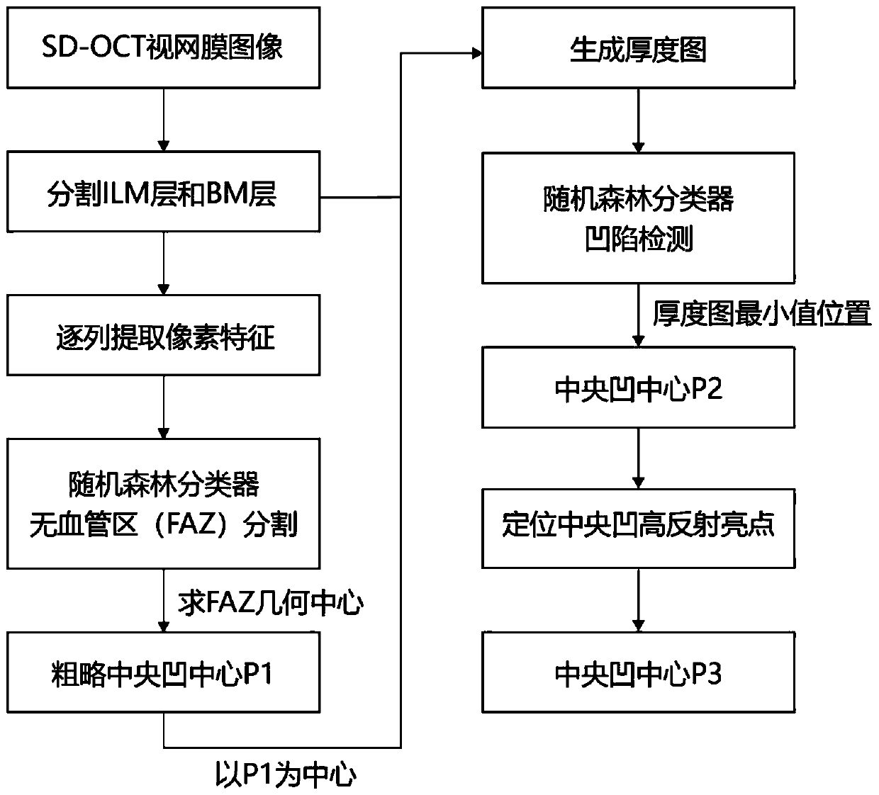
[0055] The present invention takes SD-OCT retinal three-dimensional data as input, and uses a random forest classifier combined with clinical experience to locate the central fovea of the SD-OCT retinal image.
[0056] In this embodiment, SD-OCT retinal volume data is collected by SD-OCT imaging equipment, and the pixel size of the three-dimensional data is 1024 pixels×512 pixels×128 pixels. In this embodiment, 700 SD-OCT volume data are collected, and 500 individual data are used as a training set to train a random forest model.
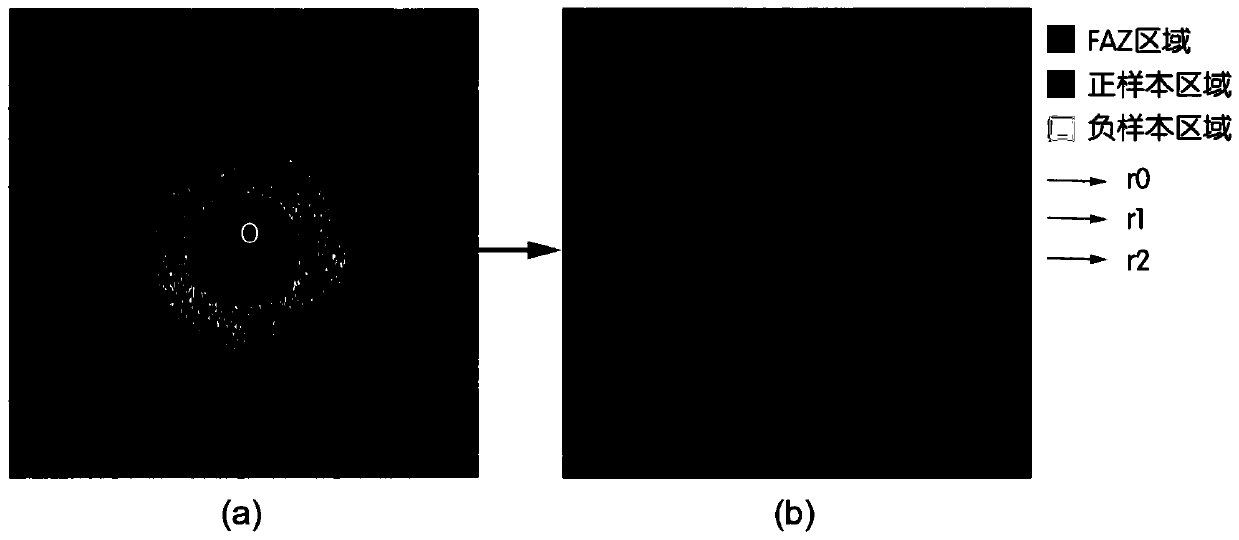
[0057] combine figure 2 , manually mark the position of the center of the macular fovea in the training data, as the gold standard, its position is recorded as O, and the avascular area is defined as a circular area with the center of the fovea O as the center and r0 = 0.25mm as the radius; O is the center of the circle, and a circular area with a radius of r1=0.15mm is used as the positive sample area, and the corresponding label value of this ...
PUM
 Abstract
Description
Claims
Application Information
 - R&D
- Intellectual Property
- Life Sciences
- Materials
- Tech Scout
- Unparalleled Data Quality
- Higher Quality Content
- 60% Fewer Hallucinations
Browse by: Latest US Patents, China's latest patents, Technical Efficacy Thesaurus, Application Domain, Technology Topic, Popular Technical Reports.
© 2025 PatSnap. All rights reserved.Legal|Privacy policy|Modern Slavery Act Transparency Statement|Sitemap|About US| Contact US: help@patsnap.com


