Single-impulse panoramic photoacoustic computed tomography (sip-pact)
A technology of light pulses and photoacoustic signals, applied in calculation, diagnosis, echo tomography, etc., can solve problems such as inability to clearly distinguish sub-organ features, poor time resolution, etc.
- Summary
- Abstract
- Description
- Claims
- Application Information
AI Technical Summary
Problems solved by technology
Method used
Image

Examples
Embodiment 1
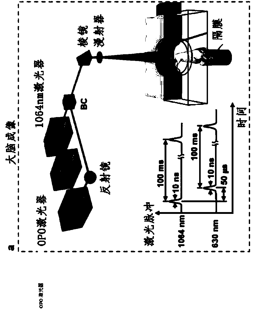
[0272] Example 1: 3D Whole Body PACT Using the SIP-PACT System
[0273] To demonstrate 3D whole-body photoacoustic computed tomography (PACT) imaging using the SIP-PACT system and method described herein, the following experiments were performed.
[0274] Adult 8-10 week-old nude mice (Hsd: Athymic Nude-FoxlNU, Harlan Co.; body weight 20-30g) were used for in vivo whole-body imaging experiments. Throughout the experiment, each mouse was maintained under anesthesia using 1.5% vaporized isoflurane. For brain imaging, mice were secured to a laboratory-made imaging platform (see Figure 25 ), and the cortical surface is positioned to align with the focal plane of the annular transducer array. During whole-body imaging experiments, the front and rear legs of the mice were strapped to the top and bottom of a laboratory-made holder, respectively, to keep the mice upright during imaging. The top of the holder consisted of an aluminum tube attached to the mouse's nose and mouth, and...
Embodiment 2
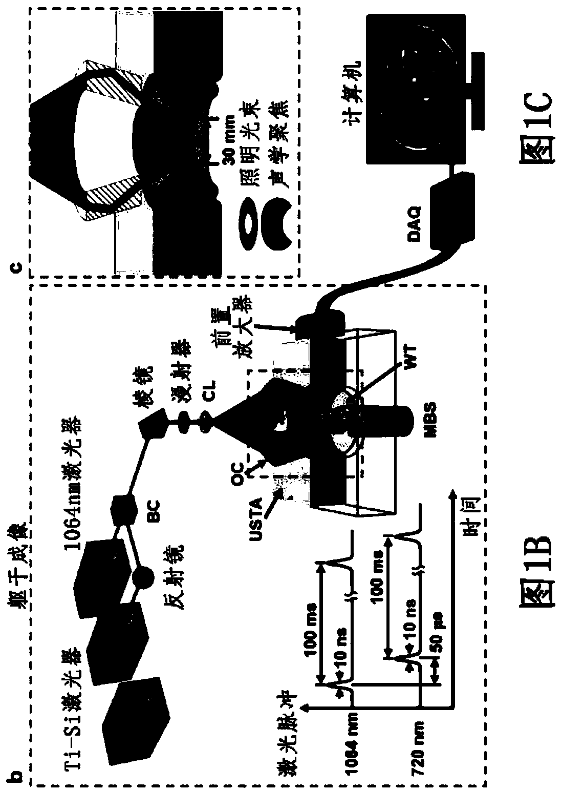
[0278] Example 2: 2D Time-Resolved Imaging of Cardiac and Respiratory Cycles Using the SIP-PACT System
[0279] To demonstrate 2D time-resolved whole-body imaging of cardiac and respiratory cycles using the SIP-PACT system and method described herein, the following experiments were performed.
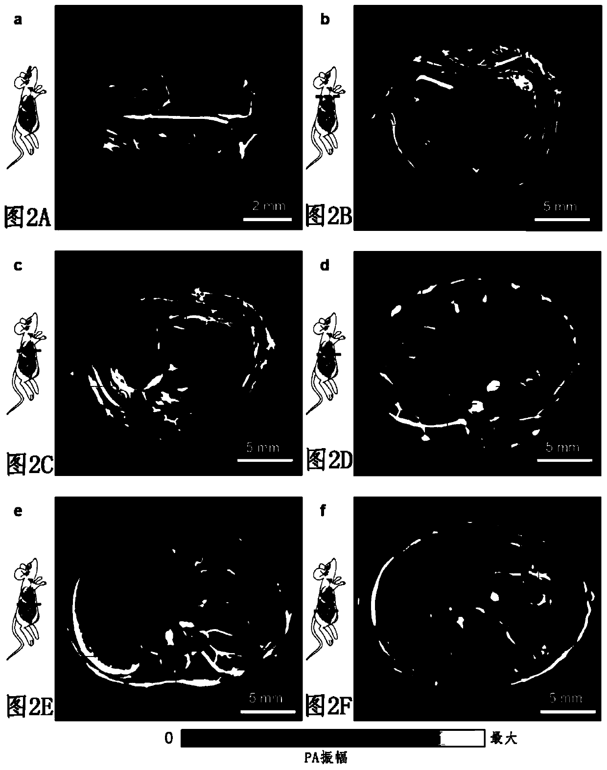
[0280] Nude mice were prepared and mounted on Figure 1B shown in the SIP-PACT system. The mouse is positioned in the SIP-PACT system such that the focal plane of the annular transducer array passes through the heart of the mouse, producing the same Figure 2B The PA image shown in is similar to the PA image. The operation of the SIP-PACT system is similar to that in Example 1, using full-circular side illumination and side detection, and emitting 1064 nm laser pulses at a repetition rate of 50 Hz to the same cross-section of the mouse thorax to obtain time-series images.
[0281] Figure 3A are representative images of transverse sections through the mouse thoracic cavity. At an i...
Embodiment 3
[0286] Example 3: Functional Imaging Using the SIP-PACT System
[0287] To demonstrate functional imaging using the SIP-PACT system and methods described herein, the following experiments were performed.
[0288] In vivo brain function and CTC imaging were performed using adult 3-4 month old Swiss Webster mice (Hsd:ND4, Swiss Webster, Harlan Co.; body weight 20-30 g). Hair was removed from each mouse with a clipper and depilatory cream before functional brain imaging using the SIP-PACT system. Each mouse was then mounted on a laboratory-fabricated imaging platform and the cortical surface of the brain was aligned with the focal plane of the transducer array as described in Example 1. Each mouse breathed inhaled gas with different oxygen concentrations to systematically adjust the oxygen saturation (sO) of hemoglobin in mice during SIP-PACT functional imaging. 2 ), as described in detail below.
[0289] Using a SIP-PACT system similar to the system described in Example 1 (se...
PUM
| Property | Measurement | Unit |
|---|---|---|
| diameter | aaaaa | aaaaa |
| distance | aaaaa | aaaaa |
| diameter | aaaaa | aaaaa |
Abstract
Description
Claims
Application Information
 - R&D
- Intellectual Property
- Life Sciences
- Materials
- Tech Scout
- Unparalleled Data Quality
- Higher Quality Content
- 60% Fewer Hallucinations
Browse by: Latest US Patents, China's latest patents, Technical Efficacy Thesaurus, Application Domain, Technology Topic, Popular Technical Reports.
© 2025 PatSnap. All rights reserved.Legal|Privacy policy|Modern Slavery Act Transparency Statement|Sitemap|About US| Contact US: help@patsnap.com