
Articular cartilage repair stent forming device and preparation method thereof

A technology of articular cartilage and molding devices, which is applied in joint implants, joint implants, additive manufacturing, etc., can solve problems affecting healing effects, errors in bracket size and wound size, etc., to improve repair effects and simplify surgical operations , Reduce the effect of cartilage loss

Inactive Publication Date: 2019-09-17
BEIJING WANJIE MEDICAL DEVICE CO LTD
View PDF12 Cites 2 Cited by
  • Summary
  • Abstract
  • Description
  • Claims
  • Application Information

AI Technical Summary

Problems solved by technology

However, since the cutting is done manually, it has a lot to do with the doctor's experience, techniques, and patience, which results in errors in the size of the obtained scaffold and the size of the wound surface, making it difficult to implant in place smoothly, seriously affecting the final healing effect

Method used


Image

  • Articular cartilage repair stent forming device and preparation method thereof
  • Articular cartilage repair stent forming device and preparation method thereof
  • Articular cartilage repair stent forming device and preparation method thereof

Examples


Embodiment 1

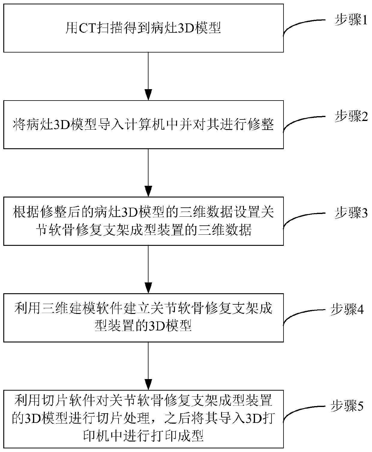
[0035] Such as figure 1 As shown, what is described in the embodiment of the present invention is a preparation method of a scaffold forming device for articular cartilage repair, the method comprising:

[0036] Step 1. Obtain the three-dimensional data of the injured part of the articular cartilage of the patient through CT examination, and obtain the 3D model of the injured articular cartilage through three-dimensional modeling software according to the obtained three-dimensional data. In this embodiment, scanning detection is performed by spiral CT to obtain three-dimensional scanning information of the lesion.

[0037] Step 2, trimming the obtained 3D model of articular cartilage damage by using a 3D modeling software, and obtaining 3D data of the trimmed 3D model of articular cartilage damage. Due to the various shapes of articular cartilage damage, often accompanied by cracks, it is necessary to properly trim the 3D model of articular cartilage damage, so that the irreg...

Embodiment 2

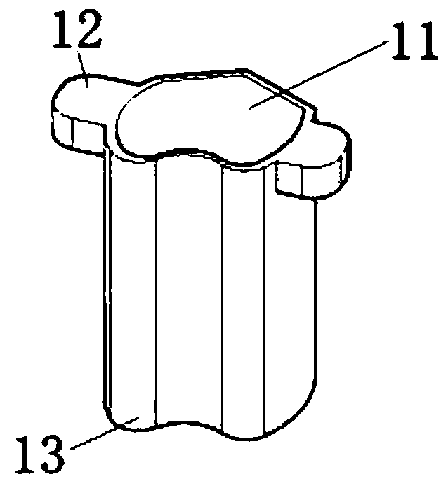
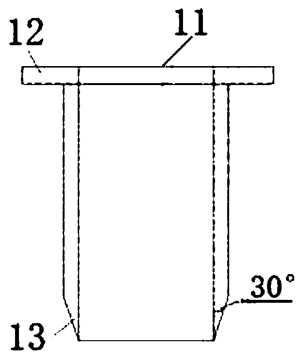
[0051] Such as Figure 2-4 As shown, the embodiment of the present invention is a scaffold forming device for articular cartilage repair, the device includes a cutter and a push rod, wherein:

[0052] The cutter is a hollow cylinder, the shape of the cutter cross section 11 is the same as that of the articular cartilage damage cross section, the size of the cutter cross section 11 is 2-5% of the overall expansion of the articular cartilage damage cross section, and the height of the cutter is 10% of the articular cartilage damage depth -20 times, the wall thickness of the cutter is 1-2mm, and the bottom end of the cutter is provided with a blade 13;

[0053] The shape of the push rod cross-section 21 is the same as that of the articular cartilage damage cross-section, the size of the push rod cross-section 21 is 2-5% of the overall reduction of the articular cartilage damage cross-section, and the height of the push rod is 1.1-1.2 times the height of the cutter;

[0054] Cutt...


PUM


Abstract

The invention discloses a preparation method of an articular cartilage repair stent forming device. The preparation method comprises the following steps: obtaining three-dimensional data of the joint cartilage injury part of a patient through CT examination, and obtaining a 3D model of the joint cartilage injury through three-dimensional modeling software; trimming the obtained 3D model of the articular cartilage injury by using the three-dimensional modeling software to obtain three-dimensional data of the trimmed articular cartilage injury 3D model; setting three-dimensional data of the articular cartilage repair stent forming device according to the three-dimensional data of the trimmed articular cartilage injury 3D model; drawing a 3D model of the articular cartilage repair stent forming device by utilizing the three-dimensional modeling software according to the three-dimensional data of the articular cartilage repair stent forming device; carrying out slicing treatment on the 3D model of the articular cartilage repair stent forming device in the step 4 by utilizing 3D printing slicing software, and then introducing the 3D model of the articular cartilage repair stent forming device subjected to slicing treatment into a 3D printer for 3D printing and forming.

Description

technical field [0001] The invention relates to the field of medical devices, in particular to a scaffold forming device for articular cartilage repair and a preparation method thereof. Background technique [0002] Human articular cartilage is an avascular tissue with no blood supply. Its nutrition comes from the moisture of the surrounding joint synovial fluid. Once damaged, it is difficult to regenerate. Therefore, cartilage repair has become one of the most challenging clinical problems for orthopedic surgeons. Cartilage damage, if not treated properly and in time, usually ends in artificial joint replacement. [0003] After decades of development, articular cartilage repair technology has gone through several eras. From the initial bone marrow stimulation technology, to the chondrocyte transplantation technology, and later the carrier that can implant the patient's chondrocytes in advance, it has developed to the current fourth-generation cartilage repair technology. ...

Claims


Application Information

Patent Timeline
IPC IPC(8): A61F2/28A61F2/46
CPCA61F2/28A61F2/2846A61F2/4644A61F2002/2835A61F2/30942A61F2002/30948A61F2002/30985A61F2002/30943
Inventor 张梅史丰田陈书香孙雪莲
Owner BEIJING WANJIE MEDICAL DEVICE CO LTD
Learn More