
Preparation method of electrochemical biosensor for simultaneously detecting HAT and TdT and application thereof

A biosensor and electrochemical technology, applied in the field of functional biomaterials and biosensing, to achieve high sensitivity, detection, and high-sensitivity detection

Active Publication Date: 2019-06-07
NINGBO UNIV
View PDF3 Cites 8 Cited by
  • Summary
  • Abstract
  • Description
  • Claims
  • Application Information

AI Technical Summary

Problems solved by technology

At present, there are no reports on electrochemical biosensors using this mechanism for HAT and TdT activity detection at home and abroad.

Method used


Image

  • Preparation method of electrochemical biosensor for simultaneously detecting HAT and TdT and application thereof
  • Preparation method of electrochemical biosensor for simultaneously detecting HAT and TdT and application thereof
  • Preparation method of electrochemical biosensor for simultaneously detecting HAT and TdT and application thereof

Examples


Embodiment 1

[0029] Example 1 Preparation of sensor

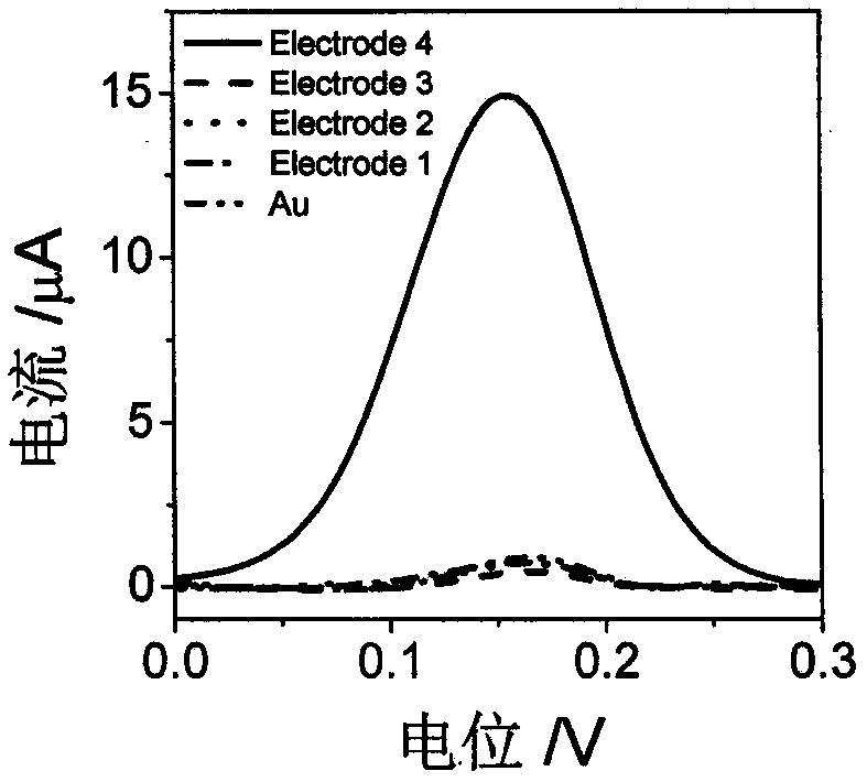
[0030] (1) Combine DNA (5μL, 0.5μM) and Hg 2+ (5μL, 1μM), mix well, put it at 37°C for 60min, and drop it on the surface of a clean bare gold electrode (Au). The electrode was slowly buffered and washed with distilled water, and then mercaptohexanol (MCH) (5 μL, 1 mM) was added dropwise and allowed to stand for 30 min at room temperature. The electrode was buffered and washed with distilled water, which was recorded as Electrode 1.

[0031] (2) Take acetyltransferase p300 (1000nM, 0.1μL), peptides (1mM, 0.4μL) and acetyl-CoA (1mM, 1μL) separately in phosphate buffer solution (PBS, 10mM, pH 7.0) and mix well, the total volume 2μL. The reaction solution was placed in a constant temperature water bath at 30°C and incubated for 30 minutes. Spread it on Electrode 1, place it at 30°C for 30 minutes, and wash the electrode slowly with distilled water, denoted as Electrode 2.

[0032] (3) Take the Exo I solution (5μL, 50U / mL) and apply it to the su...

Embodiment 2

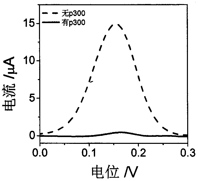
[0035] Example 2 Feasibility experiment of p300 detection

[0036] In order to prove that the sensor of the present invention can detect p300, our biosensor was prepared based on Example 1. During the acetylation reaction process in step (2), the reaction solution was used for the preparation of repair electrodes under the conditions of p300 and no p300. biological sensor. see figure 2 When there is p300, the sensor basically has no electrochemical response in PBS (0.1M, pH 7.0), but when there is no p300, there is an obvious electrochemical response, which proves that the sensor can be used for p300 activity detection.

Embodiment 3

[0037] Example 3 Detection of p300 enzyme activity

[0038] Prepare our biosensor based on Example 1 for the detection of p300 activity. The concentration of p300 is: 0, 0.01, 0.04, 0.12, 0.4, 1.2, 4, 10, 40, 100, 200, 500 nM, and the preparation of different concentrations of p300 The electrochemical response of the obtained sensor to PBS (0.1M, pH 7.0) electrolyte solution, see the experimental results image 3 , Indicating that as the concentration of p300 increases, the electrochemical response of the sensor becomes weaker, and the peak current has a good linear relationship with the logarithm of the concentration of p300. The linear correlation equation is y=8.25-3.62lgCp300, R 2 =0.9910, the linear range is 0.01~500nM, and the detection limit is 3pM, indicating that the sensor can achieve high sensitivity detection of p300 activity.


PUM


Abstract

The invention discloses a preparation method of an electrochemical biosensor for simultaneously detecting HAT and TdT and application thereof. The method comprises: step one, adding MCH dropwise ontoa hairpin DNA modified electrode surface induced by Hg<2+> and marking the processed electrode as Electrode 1; step two, taking acetyltransferase p300, polypeptide and acetylcoenzyme A respectively and mixing the acetyltransferase p300, the polypeptide and the acetylcoenzyme A fully in a phosphoric acid buffer solution (PBS, 10 mM, pH 7.0), dropping and coating the mixed solution on the surface ofthe Electrode1, and marking the obtained substance as Electrode2; step three, dropping and coating an Exo I solution on the surface of the Electrode2, carrying out incubation at a room temperature, dropping a TdT reaction solution on the electrode surface, and marking the obtained substance as Electrode3; and step four, adding Cu<2+> on the electrode surface obtained in the step three, dropping an ascorbic acid solution on the electrode surface, marking the obtained substance as Electrode 4, and detecting an electrochemical response in PBS electrolyte solution with parameters of 0.1M and pH7.0. The preparation method has the following advantages: the specificity and sensitivity are high; the detection speed is fast; the activities of two kinds of enzymes can be detected simultaneously; and the result is accurate and reliable and the cost is low.

Description

Technical field [0001] The invention relates to an electrochemical biosensor and a detection method thereof, in particular to a preparation method and application of an electrochemical biosensor for simultaneously detecting HAT and TdT, and belongs to the technical field of functional biomaterials and biosensing. Background technique [0002] Protein post-translational modification is the basis for the complex regulation and information transmission of proteins by cells. It not only affects the high-level structure and biological activity of the protein, but also regulates its subcellular location, metabolic cycle and interaction with other macromolecular substances, and the occurrence and development of cancer It is closely related to protein post-translational modification. Among them, histone acetyltransferase (HAT) mostly acts on specific lysine residues in the core histone amino acid basic amino acid concentration area. The basic principle is to transfer the acetyl group of ...

Claims


Application Information

Patent Timeline
Patent Type & Authority Applications(China)
IPC IPC(8): G01N27/327
Inventor 胡宇芳张青青胡丹丹马少华郭智勇王邃
Owner NINGBO UNIV
Learn More