
A pulmonary nodule detection method and system based on a CT image

A CT image and detection method technology, applied in image analysis, image enhancement, graphic image conversion and other directions, can solve the problems of difficult to automatically locate accurate positions, difficult to distinguish nodules, etc.

Active Publication Date: 2019-04-26
HUAZHONG UNIV OF SCI & TECH
View PDF5 Cites 31 Cited by
  • Summary
  • Abstract
  • Description
  • Claims
  • Application Information

AI Technical Summary

Problems solved by technology

Furthermore, it is also challenging to develop a robust detection system due to the high similarity between pulmonary nodules and their surrounding tissues.
For example, for concurrent nodules, since the gray scale of the nodules and the lung wall is almost the same, it is difficult for us to automatically locate its exact location
Similarly, for small nodules with a diameter of less than 6 mm, it is difficult to distinguish such nodules due to their similar gray scale with the surrounding noise

Method used


Image

  • A pulmonary nodule detection method and system based on a CT image
  • A pulmonary nodule detection method and system based on a CT image
  • A pulmonary nodule detection method and system based on a CT image

Examples


Embodiment Construction

[0053] In order to make the object, technical solution and advantages of the present invention clearer, the present invention will be further described in detail below in conjunction with the accompanying drawings and embodiments. It should be understood that the specific embodiments described here are only used to explain the present invention, not to limit the present invention. In addition, the technical features involved in the various embodiments of the present invention described below can be combined with each other as long as they do not constitute a conflict with each other.

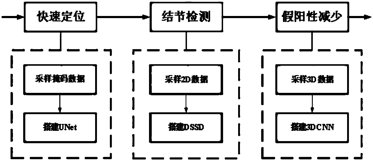
[0054] figure 1 It is an execution flowchart of the method for detecting pulmonary nodules based on CT images provided by the present invention. It includes the following steps:

[0055] 1. Quick positioning

[0056] In this step, positive and negative samples are sampled by using a boundary-based weighted sampling strategy. Note that the label of each sample here is its corresponding mask im...


PUM


Abstract

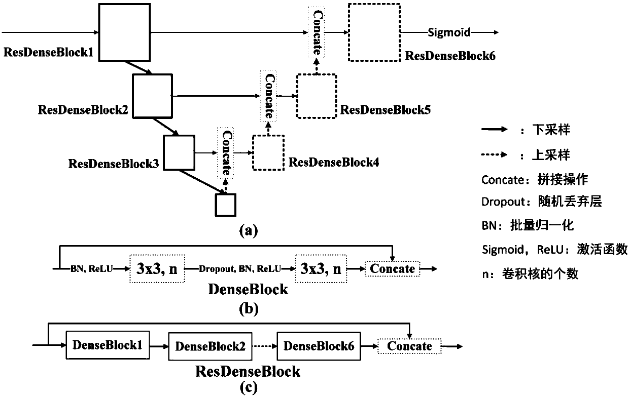
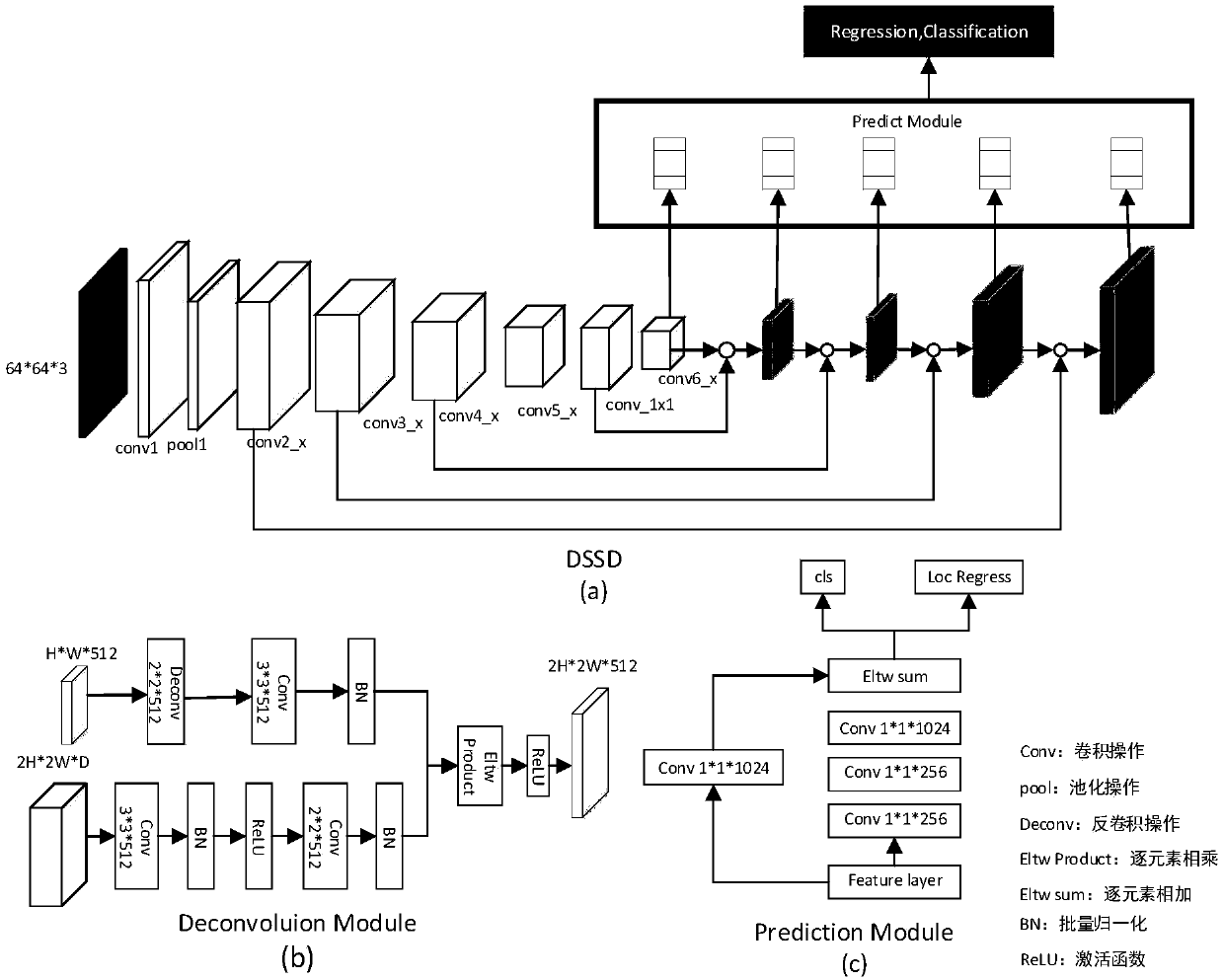
The invention discloses a pulmonary nodule detection method and system based on a CT image. The detection method specifically comprises the following steps: (1) rapid positioning processing based on aUNet network: obtaining a mask of a suspected pulmonary nodule by using a model based on the UNet network; (2) target detection processing based on a DSSD network: processing an image block corresponding to the pulmonary nodule mask obtained in the step (1) by using a DSSD network-based model to obtain a pulmonary nodule detection result; And (3) false positive screening processing based on 3DCNN: screening the candidate nodules by using a 3DCNN-based model to remove the false positive nodules. Compared with the prior art, the whole process of the method for detecting the pulmonary nodules inthe CT image and the design of all functional modules are improved, ideal detection performance is achieved, that is, the method can be used for detecting various types of pulmonary nodules, and compared with the prior art, manual intervention on a detection result can be effectively reduced.

Description

Technical field [0001] The invention belongs to the technical fields of medical image analysis and computer-aided diagnosis, and more specifically, relates to a method and system for detecting pulmonary nodules based on CT images. Background technique [0002] Lung cancer is one of the most dangerous diseases causing cancer death, accounting for two-thirds of all cancers and with a 5-year survival rate of 18%. Clinical experience shows that if lung cancer can be diagnosed at an early stage, the patient's chance of survival will be greatly improved. Diagnostic methods using lung-based computed tomography (CT) images are an important strategy for early diagnosis of lung cancer and improving patient survival rates. Among diagnostic methods based on medical imaging, accurate detection of pulmonary nodules is an important step in diagnosing early lung cancer. It is of great clinical significance to develop a robust automatic detection system for pulmonary nodules. However, due...

Claims


Application Information

Patent Timeline
IPC IPC(8): G06T7/00G06T7/136G06T3/40
CPCG06T3/4038G06T7/0012G06T2207/10081G06T2207/20081G06T2207/20084G06T2207/20221G06T2207/30064G06T7/136
Inventor 刘宏曹海潮马光志宋恩民金勇刘楚华刘腾营金人超许向阳
Owner HUAZHONG UNIV OF SCI & TECH
Learn More