
Eye fundus image splicing method and system

A fundus image, image registration technology, applied in the field of medical image processing

Active Publication Date: 2019-04-19
代黎明
View PDF2 Cites 10 Cited by
  • Summary
  • Abstract
  • Description
  • Claims
  • Application Information

AI Technical Summary

Problems solved by technology

[0005] Based on this, it is necessary to provide a fundus image stitching method and system for the problem that the traditional fundus image stitching method has certain limitations.

Method used


Image

  • Eye fundus image splicing method and system
  • Eye fundus image splicing method and system
  • Eye fundus image splicing method and system

Examples


Embodiment Construction

[0064] In order to make the technical solution of the present invention clearer, the present invention will be further described in detail below in conjunction with specific examples. Wherein, it should be noted that the following description includes various specific details to facilitate understanding, but these details are to be regarded as merely exemplary. Accordingly, those of ordinary skill in the art will recognize that various changes and modifications of the various embodiments described herein can be made without departing from the scope and spirit of the disclosure. Also, descriptions of well-known functions and constructions may be omitted for clarity and conciseness.

[0065] The terms and words used in the following description and claims are not limited to the bibliographical meanings, but, are merely used by the inventor to enable a clear and consistent understanding of the present disclosure. Accordingly, it should be apparent to those skilled in the art tha...


PUM


Abstract

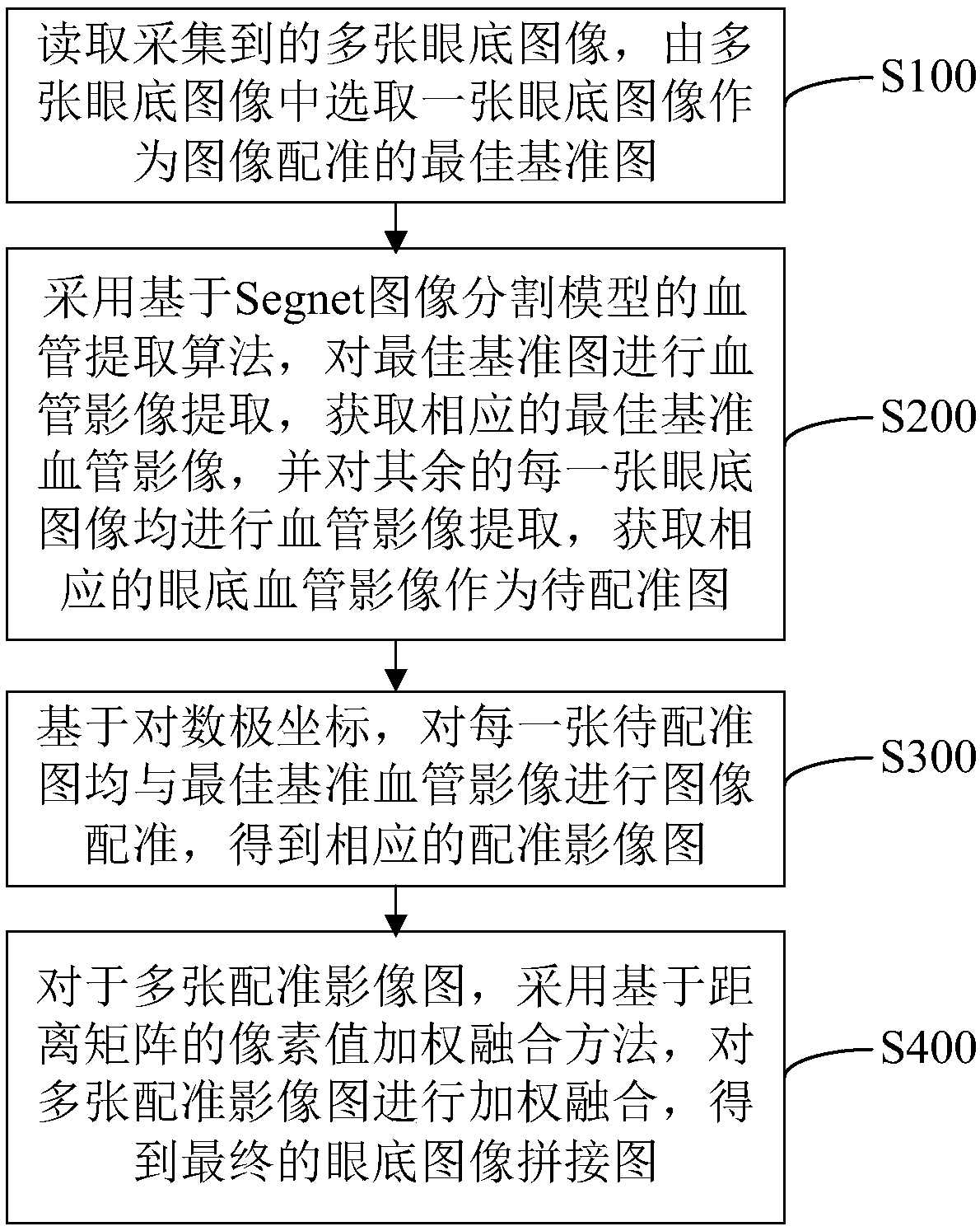
The invention discloses an eye fundus image splicing method and system, and the method comprises the steps: reading a plurality of collected eye fundus images, and selecting one eye fundus image fromthe plurality of eye fundus images as an optimal reference image for image registration; Carrying out blood vessel image extraction on the optimal reference image by adopting a blood vessel extractionalgorithm based on a Segnet image segmentation model, obtaining a corresponding optimal reference blood vessel image, carrying out blood vessel image extraction on each of the rest eye fundus images,and obtaining a corresponding eye fundus blood vessel image as a to-be-registered image; Based on the logarithmic polar coordinates, performing image registration on each to-be-registered image and the optimal reference blood vessel image to obtain a corresponding registered image; And for the plurality of registered image images, carrying out weighted fusion on the plurality of registered imageimages by adopting a pixel value weighted fusion method based on a distance matrix to obtain a final fundus image splicing image. The quality of the stitched image is effectively improved, and the problem that a traditional fundus image stitching method has a certain limitation is solved.

Description

technical field [0001] The invention relates to the technical field of medical image processing, in particular to a fundus image stitching method and system. Background technique [0002] Neonatal fundus images are an objective and standard auxiliary diagnostic method in neonatal ophthalmology. As the only blood vessels that can be directly observed in the whole body, retinal blood vessels are effective indicators for evaluating the microvascular function of the whole body, and the inspection method is simple and noninvasive. Therefore, the fundus images of newborns can not only record the eye conditions of newborns at birth, but also facilitate remote consultation and preserve evidence-based medical evidence, providing a wealth of information for newborn eye disease screening, retinal disease diagnosis and treatment, and pathological mechanism exploration. Information. In the process of taking fundus images, because the color of the retina of the fundus of newborns is ligh...

Claims


Application Information

Patent Timeline
IPC IPC(8): G06T7/00G06T7/33G06T7/11G06T7/136G06T3/40
CPCG06T3/4038G06T7/0012G06T7/11G06T7/136G06T7/33G06T2207/30101G06T2207/30041G06T2207/10004
Inventor 高孟娣姜泓羊杨康代黎明
Owner 代黎明
Learn More