Preparation method and application of a kind of keratitis animal model
An animal model, keratitis technology, applied in the field of animal models, can solve the problems of complex surgery, many complications and other factors
- Summary
- Abstract
- Description
- Claims
- Application Information
AI Technical Summary
Problems solved by technology
Method used
Image
Examples
Embodiment 1
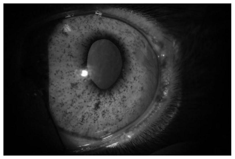
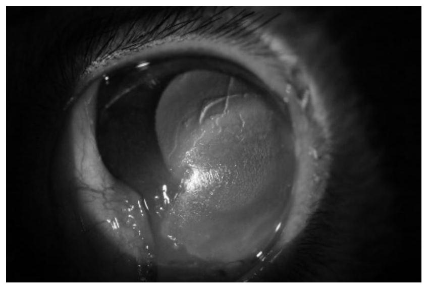
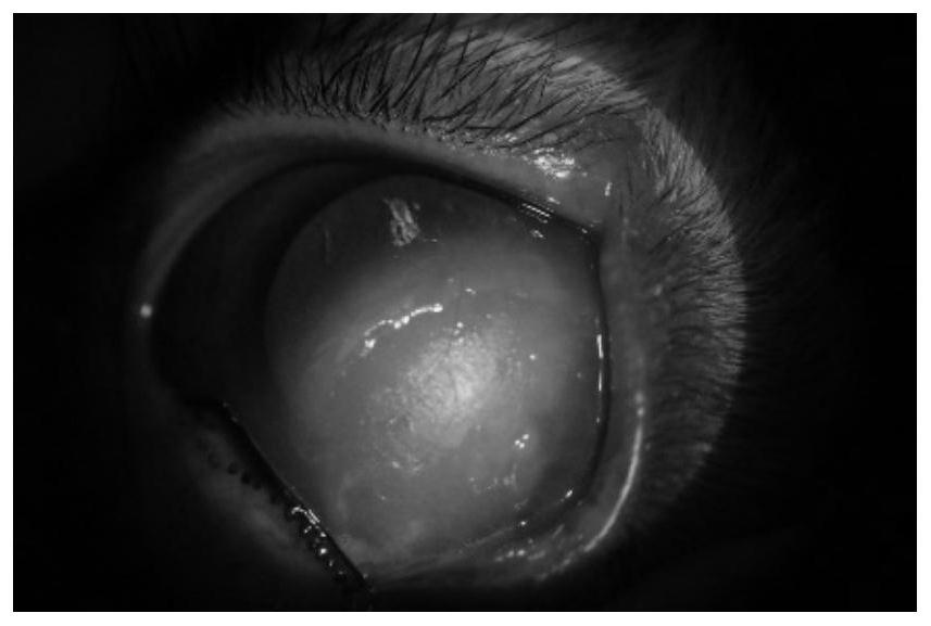
[0073] The embodiment of the present application provides a method for preparing an animal model of keratitis.
[0074] 1. Exposure of the ophthalmic branch of the trigeminal nerve
[0075] a, anesthesia: weigh the quality of the male cyanotic blue rabbit to be 2.6kg, give the rabbit intravenous injection of 5.2mL of 1% pentobarbital sodium solution;
[0076] b. Opening: press the zygomatic fossa at the corner of the rabbit’s eye to slightly protrude the eyeball to expose the rectus oculi muscle, and use ophthalmic scissors to pull the fascia at the fornix between the lateral rectus muscle and the inferior rectus muscle on the lower edge of the rabbit’s eyeball to form an opening;
[0077] c. Remove obstacles: Use ophthalmic forceps to bluntly separate the fascia between the lateral rectus muscle and the inferior rectus muscle to make the lacrimal gland visible, and gently pull the lacrimal gland to the outside of the rabbit’s eyeball to make the inner canthal venous plexus in...
Embodiment 2
[0084] The embodiment of the present application provides a method for preparing an animal model of keratitis.
[0085] 1. Exposure of the ophthalmic branch of the trigeminal nerve
[0086] a, anesthesia: weigh the quality of the male cyanotic blue rabbit to be 2.4kg, inject 4.3mL of pentobarbital sodium solution with a mass fraction of 0.12% to the rabbit;
[0087] b. Opening: press the zygomatic fossa at the corner of the rabbit’s eye to slightly protrude the eyeball to expose the rectus oculi muscle, and use micro-scissors to pull the fascia at the fornix between the lateral rectus muscle and the inferior rectus muscle on the lower edge of the rabbit’s eyeball to form an opening;
[0088] c. Remove obstacles: Use micro-tweezers to bluntly separate the fascia between the lateral rectus muscle and the inferior rectus muscle to make the lacrimal gland visible, and gently pull the lacrimal gland to the outside of the rabbit’s eyeball to make the inner canthal venous plexus insi...
Embodiment 3
[0095] The embodiment of the present application provides a method for preparing an animal model of keratitis.
[0096] 1. Exposure of the ophthalmic branch of the trigeminal nerve
[0097] a, anesthesia: weigh the quality of the male cyanotic blue rabbit to be 2.7kg, inject 5.9mL of pentobarbital sodium solution with a mass fraction of 0.08% to the rabbit;
[0098] b. Opening: press the zygomatic fossa at the corner of the rabbit’s eye to slightly protrude the eyeball to expose the rectus oculi muscle, and use micro-scissors to pull the fascia at the fornix between the lateral rectus muscle and the inferior rectus muscle on the lower edge of the rabbit’s eyeball to form an opening;
[0099] c. Remove obstacles: Use micro-tweezers to bluntly separate the fascia between the lateral rectus muscle and the inferior rectus muscle to make the lacrimal gland visible, and gently pull the lacrimal gland to the outside of the rabbit’s eyeball to make the inner canthal venous plexus insi...
PUM
 Abstract
Description
Claims
Application Information
 - R&D
- Intellectual Property
- Life Sciences
- Materials
- Tech Scout
- Unparalleled Data Quality
- Higher Quality Content
- 60% Fewer Hallucinations
Browse by: Latest US Patents, China's latest patents, Technical Efficacy Thesaurus, Application Domain, Technology Topic, Popular Technical Reports.
© 2025 PatSnap. All rights reserved.Legal|Privacy policy|Modern Slavery Act Transparency Statement|Sitemap|About US| Contact US: help@patsnap.com


