X-ray source array, x-ray tomography system and method
An X-ray and array technology, applied in the field of X-ray scanning imaging, can solve the problems of different magnification, large distance difference, unfavorable system integration, etc., and achieve the effect of reducing volume and improving quality
- Summary
- Abstract
- Description
- Claims
- Application Information
AI Technical Summary
Problems solved by technology
Method used
Image
Examples
Embodiment 1
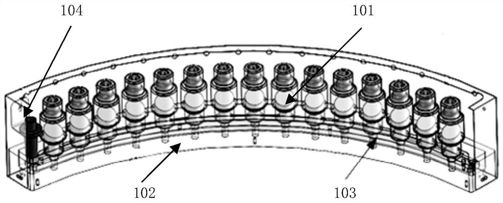
[0029] The technical solution of this embodiment is applicable to the field of X-ray tomographic imaging. The X-ray source array of the embodiment of the present invention may include a plurality of individually packaged cold cathode X-ray source bulbs; wherein each cold cathode X-ray source bulb The arrangement track of the tubes is arc-shaped.
[0030] Among them, the cold cathode X-ray source tube adopts the cold cathode field emission X-ray source, which generates electron beams by means of field electron emission. The field emission cathode has low operating temperature, low power consumption, and is easy to integrate. At the same time, since there is no time delay in the field electron emission, the field emission X-ray source using a cold cathode can achieve high time resolution and programmable X-ray emission. It can be understood that the X-ray source array may include multiple cold-cathode X-ray source tubes. Optionally, the number of cold-cathode X-ray source tubes ...
Embodiment 2
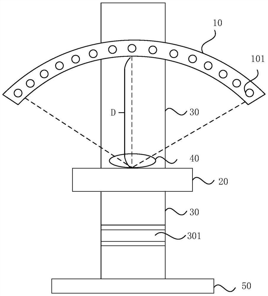
[0041] The technical solution of this embodiment can be applied to the field of X-ray tomography imaging, and the X-ray tomography system provided by this embodiment includes a detector and any X-ray source array of the above-mentioned embodiments; wherein, the arrangement of the X-ray source array The center of the circle where the trajectory is located is on the detector. The principle and specific implementation of the X-ray source array are as described in the above embodiments.
[0042] Among them, the detector is a device that converts X-ray energy into electrical signals that can be recorded. The detector is located below the X-ray source array. After receiving the X-rays passing through the scanned object placed above the detector, it generates electrical signals proportional to the radiation intensity of the X-rays to obtain projection images from different angles.
[0043] According to the technical solution of the embodiment of the present invention, the center of ...
Embodiment 3
[0050] This embodiment is applicable to the case of X-ray static tomography, especially suitable for the case where the tube currents of each cold-cathode X-ray source tube are different and the pulse exposure time is short. The method can be executed by the X-ray tomographic scanning system provided in the second embodiment of the present invention, and the system can be realized by software and / or hardware. The method of the embodiment of the present invention specifically includes the following steps:
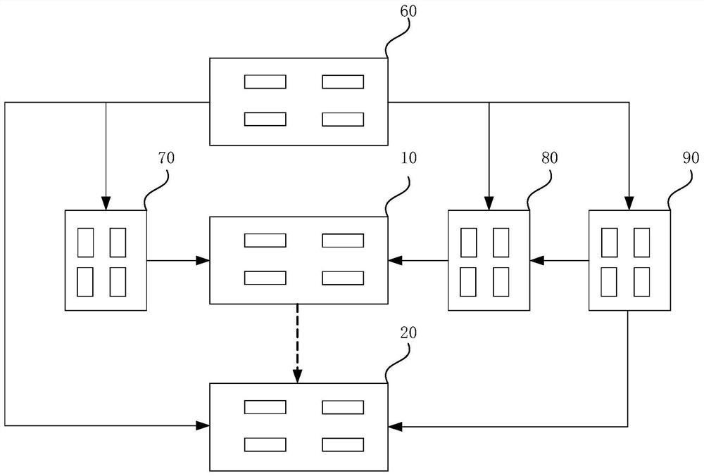
[0051] Separately calibrate the operating voltage of each cold cathode X-ray source tube in the X-ray source array, so that the tube current of each cold cathode X-ray source tube is consistent; the X-ray source array includes a plurality of individually packaged cold cathode X-ray sources The tube; scan the target object according to the operating voltage of each cold cathode X-ray source tube and the preset point-by-point pulse exposure method; control the control platform...
PUM
 Abstract
Description
Claims
Application Information
 - R&D
- Intellectual Property
- Life Sciences
- Materials
- Tech Scout
- Unparalleled Data Quality
- Higher Quality Content
- 60% Fewer Hallucinations
Browse by: Latest US Patents, China's latest patents, Technical Efficacy Thesaurus, Application Domain, Technology Topic, Popular Technical Reports.
© 2025 PatSnap. All rights reserved.Legal|Privacy policy|Modern Slavery Act Transparency Statement|Sitemap|About US| Contact US: help@patsnap.com


