RT-PCR detection primers and detection method of subgroup k avian leukosis virus
An avian leukemia virus, RT-PCR technology is applied to the K subgroup avian leukemia virus RT-PCR detection primer and detection field, which can solve the problems of easy contamination, high cost of detection reagents and consumables, inconvenience for promotion and application on breeding sites, and achieves specificity. The effect of high performance, low detection cost, and easy configuration
- Summary
- Abstract
- Description
- Claims
- Application Information
AI Technical Summary
Problems solved by technology
Method used
Image
Examples
Embodiment 1
[0026] Embodiment 1 K subgroup avian leukosis virus RT-PCR detection primer and detection method of the present invention
[0027] 1 test material
[0028] 1.1 Main reagents and instruments
[0029] AxyPrep Body Fluid Viral DNA / RNA Mini Kit (Cat No.: AP-MN-BF-VNA-250), TaKaRaPrimeScript 0ne step RT-PCR Kit Ver.2 (TaKaRa Code: DRR055A), Thermo High Speed Low Temperature Centrifuge, Thermo PCR instrument, electrophoresis instrument.
[0030] 1.2 Strains
[0031] Clinically identified avian leukosis virus by virus isolation and ELISA detection, clinically identified avian leukosis virus of A, B, J and K subgroups.
[0032] 1.3 Primer design
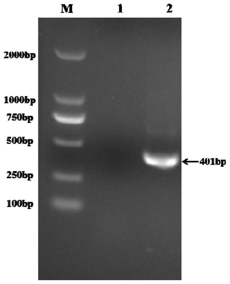
[0033] Design a pair of primers according to the env gene sequence of K subgroup virus GD14LZ strain (GenBank: KU605774.1) as shown in the table below to amplify part of the gene fragment. The expected size of the target band is 401bp. If the target fragment is amplified, it can be determined as subgroup K avian leukemia virus.
[00...
Embodiment 2
[0046] Example 2 specificity verification
[0047] Samples were used: negative control; samples positive for p27 detected by ELISA in production; subgroup K virus as positive control; subgroup A, B, and J viruses.
[0048] Test method: According to the detection method in Example 1, each sample was amplified respectively, and identified and compared by agarose gel electrophoresis respectively.
[0049] Test results: In the samples that were detected as p27 positive by ELISA in production, the K subgroup virus could amplify the target fragment, and the A, B, J subgroup viruses and other subgroup viruses isolated in production could not amplify the target fragment ( like figure 2 ).
Embodiment 3
[0050] Example 3 Sensitivity Verification
[0051] Samples used: negative control; K subgroup avian leukosis virus.
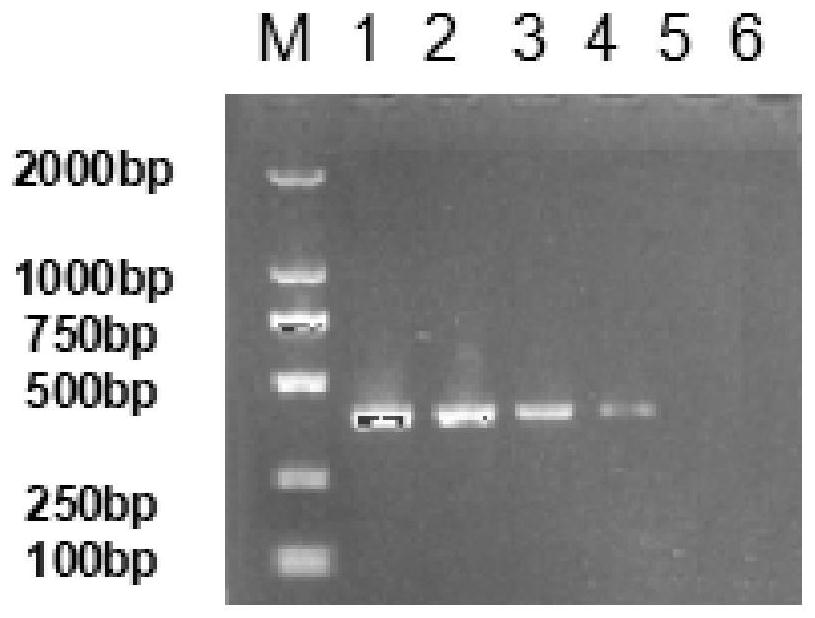
[0052] Test method: extract K subgroup avian leukosis virus sample RNA according to the detection method of embodiment 1, measure RNA concentration, and RNA is carried out 10 times ratio dilutions, each dilution RNA is amplified respectively, and respectively through agarose coagulation Gel electrophoresis identification comparison.
[0053] For test results, see image 3 : The sample RNA concentration is 95.07ng / μl, and the sample is undiluted and 10 -1 、10 -2 、10 -3 The target fragment can be amplified at all dilutions, and the concentration of the amplified product decreases sequentially. samples in 10 -4 The dilution cannot amplify the target fragment.
PUM
 Abstract
Description
Claims
Application Information
 - R&D Engineer
- R&D Manager
- IP Professional
- Industry Leading Data Capabilities
- Powerful AI technology
- Patent DNA Extraction
Browse by: Latest US Patents, China's latest patents, Technical Efficacy Thesaurus, Application Domain, Technology Topic, Popular Technical Reports.
© 2024 PatSnap. All rights reserved.Legal|Privacy policy|Modern Slavery Act Transparency Statement|Sitemap|About US| Contact US: help@patsnap.com


