A cancer cell separation device based on cell deformation quantity and dielectrophoresis force and a control system
A separation device and dielectrophoretic force technology, applied in the field of cell sorting and separation, can solve problems such as inability to separate cells
- Summary
- Abstract
- Description
- Claims
- Application Information
AI Technical Summary
Problems solved by technology
Method used
Image

Examples
preparation example Construction
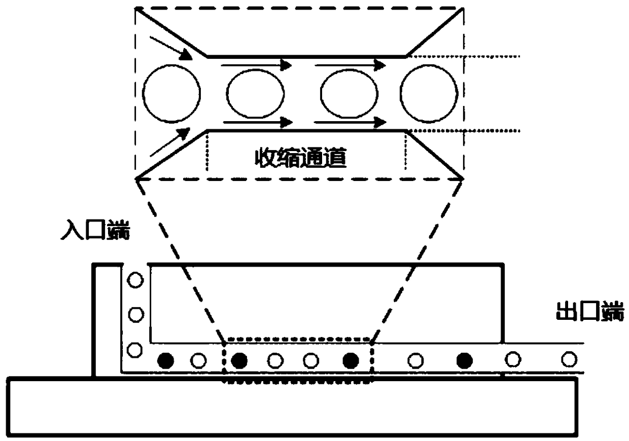
[0047] The fabrication process of the microchannels is as follows: the microchannels are created on top of the electrodes by building a 20 μm thick layer of SU-8 using standard photolithography techniques.
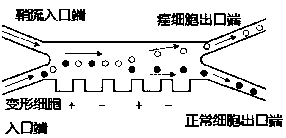
[0048] On one side of the inner wall of the microchannel, there are a plurality of electrodes with a tooth-like structure embedded, and the positive and negative electrodes are intersected to generate dielectrophoretic force in the microchannel; the dielectrophoretic force generated by the electrodes acts on the microchannel amebocytes.
[0049] The electrode on the wall of the microchannel of the cancer cell separation chip 5 is platinum metal, and platinum is a kind of inert metal, which itself does not participate in the electrode reaction when it is used as an electrode.
[0050] In order not to damage the cells, a porous hydrogel layer was laid on the platinum electrodes, which can avoid the contact between the deformed cells and the electrodes.
[0051] The electrod...
Embodiment 2
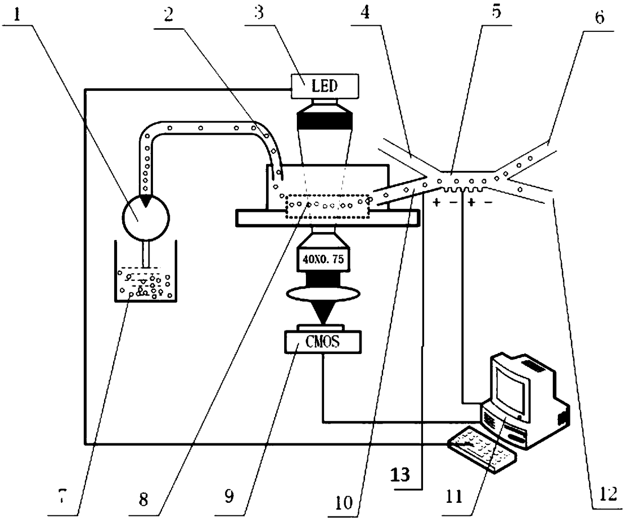
[0057] An embodiment of the present invention provides a cancer cell separation control system based on cell deformation. The cancer cell separation control system is applied to the device provided in Embodiment 1. The cancer cell separation control system includes: the CMOS high-speed camera 9 It is used to capture the image of the deformed cells in the cell deformation detection chip 8 .
[0058] The processor 11 includes a processing module and a judging module.
[0059] The processing module is connected with the CMOS high-speed camera 9, and is used for acquiring the image of the amebocyte, and processing the image of the amebocyte to determine the amount of deformation of the amebocyte.
[0060] The judging module, connected to the electrode, is used to judge whether the deformation of the deformed cells is greater than a preset threshold stored in the processor, and when the deformation of the deformed cells is greater than the preset threshold, When the drive signal i...
Embodiment 3
[0062] The embodiment of the present invention provides a method for measuring the deformation of cancer cells and separating them based on dielectrophoretic force technology. The specific implementation steps are as follows:
[0063] a. Preparation of cells: prepare the blood samples to be tested in advance, centrifuge the samples at 115g for 5min, and then resuspend them in phosphate buffered saline (PBS) so that the final concentration of the cells is 10 6 cells / mL, only 100 μL of cell suspension is required for each detection. Prepare an isotonic solution so that the conductivity of the cell suspension is 0.55 S / m during the experiment. Before aspirating the cell suspension from the cell reservoir 7 into 1 mL of the syringe pump 1, the cell suspension was placed at 37° C. to maintain cell viability.
[0064] b. Experimental device preparation: thoroughly clean the channels of the syringe micropump 1, the polymer tube 2, the cell deformation detection chip 8, and the cell ...
PUM
| Property | Measurement | Unit |
|---|---|---|
| length | aaaaa | aaaaa |
| width | aaaaa | aaaaa |
| width | aaaaa | aaaaa |
Abstract
Description
Claims
Application Information
 - R&D
- Intellectual Property
- Life Sciences
- Materials
- Tech Scout
- Unparalleled Data Quality
- Higher Quality Content
- 60% Fewer Hallucinations
Browse by: Latest US Patents, China's latest patents, Technical Efficacy Thesaurus, Application Domain, Technology Topic, Popular Technical Reports.
© 2025 PatSnap. All rights reserved.Legal|Privacy policy|Modern Slavery Act Transparency Statement|Sitemap|About US| Contact US: help@patsnap.com