CT image-based lung segmentation method, device and computer-readable storage medium

A CT image and image technology, applied in the field of CT image-based lung segmentation and computer-readable storage media, can solve problems such as loss of accuracy, smooth lung edges, and inability to effectively remove CT image noise, so as to ensure integrity, The effect of avoiding missed diagnosis

Active Publication Date: 2021-03-30
GUANGZHOU UNIVERSITY
View PDF5 Cites 0 Cited by
  • Summary
  • Abstract
  • Description
  • Claims
  • Application Information

AI Technical Summary

Problems solved by technology

However, during the research and practice of the prior art, the inventors of the present invention found that the existing lung segmentation techniques are of various types, which are not only messy but also unable to effectively remove the noise in the CT image, and will make the edges of the lungs too smooth and distorted. loss of precision

Method used


Image

  • CT image-based lung segmentation method, device and computer-readable storage medium
  • CT image-based lung segmentation method, device and computer-readable storage medium
  • CT image-based lung segmentation method, device and computer-readable storage medium

Examples


no. 1 example

[0048] see Figure 1-9 .

[0049] like figure 1 As shown, the CT image-based lung segmentation method provided in this embodiment is suitable for execution in a computer device, and at least includes the following steps:

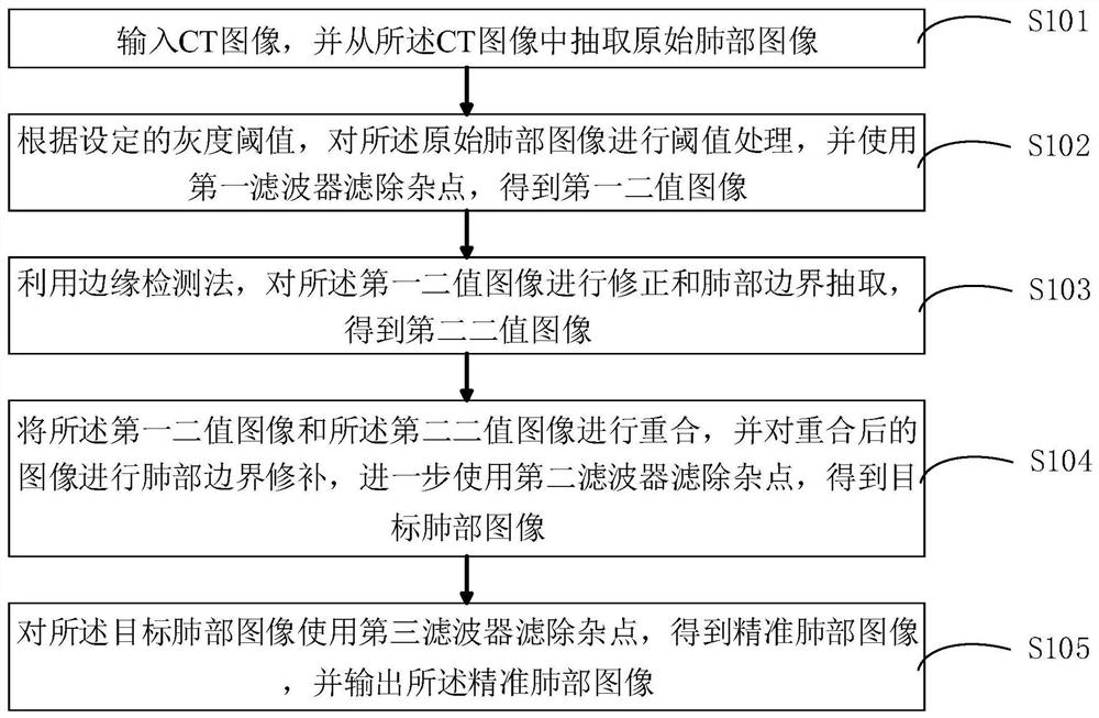
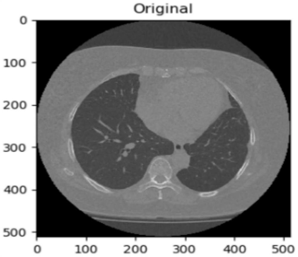
[0050] S101. Input a CT image, and extract an original lung image from the CT image;

[0051] S102. Perform threshold processing on the original lung image according to the set grayscale threshold, and use a first filter to filter out noise points to obtain a first binary image;

[0052] S103. Using an edge detection method, perform correction and lung boundary extraction on the first binary image to obtain a second binary image;

[0053] S104. Superimpose the first binary image and the second binary image, and perform lung boundary repair on the superimposed image, and further use a second filter to filter out noise points to obtain a target lung image;

[0054] S105. Use a third filter to filter out noise on the target lung image to obtain an accurate ...

no. 2 example

[0082] see Figure 10 .

[0083] like Figure 10 As shown, the present embodiment also provides a lung segmentation device based on CT images, including:

[0084] The original lung image extraction module 201 is configured to input a CT image and extract an original lung image from the CT image.

[0085] The threshold processing module 202 is configured to perform threshold processing on the original lung image according to the set grayscale threshold, and use a first filter to filter out noise points to obtain a first binary image.

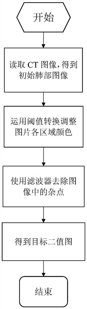
[0086] Specifically, according to the set grayscale threshold, convert the tissues other than the lungs in the original lung image into white, convert the lung tissue and air into black, and use the first filter to filter out the black parts in the image The white noise points of the white part and the black noise points of the white part are obtained to obtain the first binary image.

[0087] Wherein, the grayscale threshold is -500, and the...


PUM


Abstract

The invention discloses a lung segmentation method based on a CT image, a device and a computer-readable storage medium. The method comprises: inputting a CT image and abstracting the original image of a lung; according to the set gray threshold, performing threshold processing on the original lung image, and using a first filter to filter out impurity points to obtain a first binary image; the first binary image being modified and the lung boundary being extracted by edge detection to obtain the second binary image. The first binary image and the second binary image are coincident, the coincident image is repaired by lung boundary, and the impurity points are further filtered by a second filter to obtain a target lung image; a third filter is used to filter out impurity points to obtain an accurate lung image and output the accurate lung image. The invention can automatically perform accurate lung segmentation on a CT image, ensure the integrity of lung parenchyma region segmentation,and avoid the problem of missed diagnosis in the subsequent diagnosis process due to the loss of the edge and the region of the lung region.

Description

technical field [0001] The present invention relates to the technical field of image processing, in particular to a CT image-based lung segmentation method, device and computer-readable storage medium. Background technique [0002] In the past 50 years, the incidence of lung cancer has increased significantly. In industrialized countries in Europe and the United States and some large industrial cities in my country, the incidence of lung cancer has ranked first among malignant tumors in men, and the incidence of lung cancer has also increased rapidly in women. The second or third place has become a major disease that endangers life and health. Therefore, it is very important to improve the computer diagnosis and treatment system for lung cancer. [0003] With the development of computer technology, CT detection technology and image processing technology, the current CT image has the characteristics of high definition and high contrast, and lung segmentation technology is an ...

Claims


Application Information

Patent Timeline
Patent Type & Authority Patents(China)
IPC IPC(8): G06T7/12G06T7/13G06T7/136G06T5/10G06T5/00
CPCG06T5/002G06T5/10G06T2207/10081G06T2207/30061G06T7/12G06T7/13G06T7/136
Inventor 黄文恺薛义豪胡凌恺倪皓舟彭广龙何杰贤朱静吴羽
Owner GUANGZHOU UNIVERSITY
Why Eureka
Try Eureka