
Construction method and application of aortic dissection segmentation model

A technology for aortic dissection and segmentation models, which is applied in the field of medical imaging and can solve the problems of unpublished related work on dissection segmentation

Active Publication Date: 2018-11-13
HUIYING MEDICAL TECH (BEIJING) CO LTD
View PDF4 Cites 14 Cited by
  • Summary
  • Abstract
  • Description
  • Claims
  • Application Information

AI Technical Summary

Problems solved by technology

However, there is no published related work on the segmentation of dissections (true and false lumens)

Method used


Image

  • Construction method and application of aortic dissection segmentation model
  • Construction method and application of aortic dissection segmentation model
  • Construction method and application of aortic dissection segmentation model

Examples


Embodiment 1

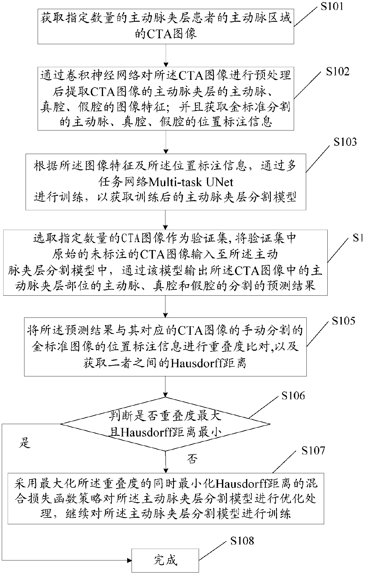
[0057] like figure 1 Shown, for the construction method of a kind of aortic dissection segmentation model that the application provides, comprise:

[0058] S101. Acquire CTA images of aortic regions of a specified number of patients with aortic dissection. The CTA image may be obtained from a large number of existing CTA images of the aortic region of patients with aortic dissection.

[0059] S102, preprocessing the CTA image through a convolutional neural network, and extracting the image features of the aorta, true lumen, and false lumen of aortic dissection in the preprocessed CTA image; and obtaining the golden standard segmented aorta Position labeling information of artery, true lumen and false lumen.

[0060] Wherein, the preprocessing includes:

[0061] Normalize the image resolution so that the x, y, and z axis resolutions are all 1mm;

[0062] Convert the image pixel value into a Hu value, and limit the Hu value in the range of (0, 600), and normalize the image H...

Embodiment 2

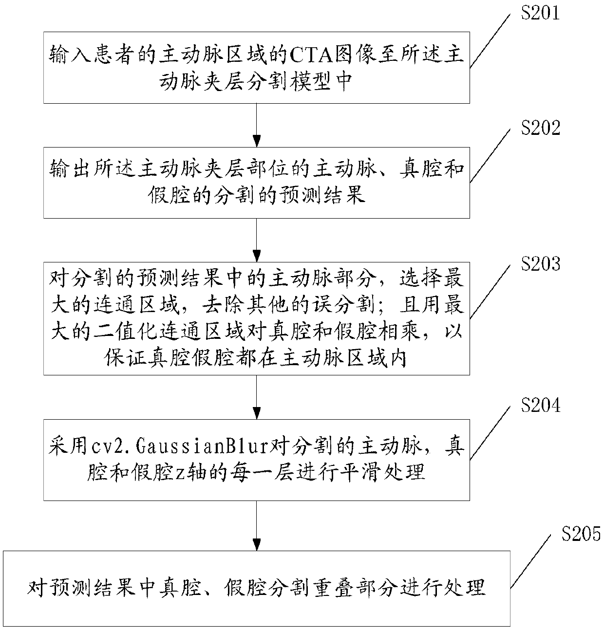
[0096] The present application also provides a method for aortic dissection segmentation based on the above-mentioned aortic dissection segmentation model, comprising the following steps:

[0097] S201, inputting the CTA image of the aortic region of the patient into the aortic dissection segmentation model;

[0098] S202. Outputting a prediction result of segmentation of the aorta, true lumen, and false lumen at the aortic dissection site.

[0099] After the step S202, post-processing of the segmented prediction results is also included, specifically:

[0100] S203, select the largest connected area for the aorta part in the segmentation prediction result, and remove other mis-segmentation; and multiply the true lumen and the false lumen by the largest binary connected area to ensure that the true lumen and the false lumen are all in within the aortic region; and / or

[0101] S204, using cv2.GaussianBlur to smooth each layer of the segmented aorta, true lumen and false lumen...


PUM


Abstract

The invention provides a construction method and application of an aortic dissection segmentation model. The method comprises the following steps: A. acquiring a specified number of CTA images of aortic regions of aortic dissection patients; B, preprocessing the CTA images through a convolutional neural network, and extracting image features of the aorta, a true cavity and a pseudo-cavity of the aortic dissection of the preprocessed CTA images, and acquiring location annotation information of the aorta, the true cavity and the pseudo-cavity divided by the golden standard; and C, performing training according to the image features and the location annotation information through a multi-task network Multi-task UNet to obtain a trained aortic dissection segmentation model. Therefore, a segmentation prediction result of the aortic dissection can be obtained quickly and effectively via the above model, the diagnosis time of a doctor is greatly shortened, and an effective support is providedfor the surgical plan formulation.

Description

technical field [0001] The invention relates to the field of medical imaging, in particular to a construction method and application of an aortic dissection segmentation model. Background technique [0002] The treatment of aortic dissection is usually accomplished by transplanting a covered stent. Before the operation, the doctor needs to make a prognosis and determine a specific surgical plan based on the morphological parameters of the dissection (such as the largest diameter of the true cavity), such as selecting a stent of an appropriate size. After the operation, doctors also need to judge the effect of the operation according to the morphological parameters of the dissection. The morphological parameters of the dissection (such as the diameter of true and false lumens) can be obtained by segmenting the aortic dissection. At present, the segmentation methods of aortic dissection are mainly divided into traditional segmentation methods. Traditional methods mainly incl...

Claims


Application Information

Patent Timeline
IPC IPC(8): G06K9/34G06K9/62
CPCG06V10/267G06V2201/03G06F18/22
Inventor 柴象飞郭伟郭娜葛阳阳左盼莉曹龙孟博文王成李健宁
Owner HUIYING MEDICAL TECH (BEIJING) CO LTD
Learn More