
MRI (Magnetic Resonance Imaging) brain tumor localization and intratumoral segmentation method based on deep cascaded convolution network

A convolutional network and tumor localization technology, which is applied in the field of MRI brain tumor localization and intratumor segmentation based on deep convolutional convolutional network, which can solve the problems of inaccurate segmentation boundaries of small areas, and the segmentation speed is not utilized by the image block classification method.

Active Publication Date: 2018-09-04
CHONGQING NORMAL UNIVERSITY
View PDF11 Cites 70 Cited by
  • Summary
  • Abstract
  • Description
  • Claims
  • Application Information

AI Technical Summary

Problems solved by technology

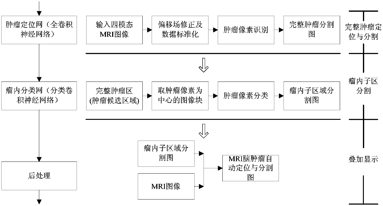
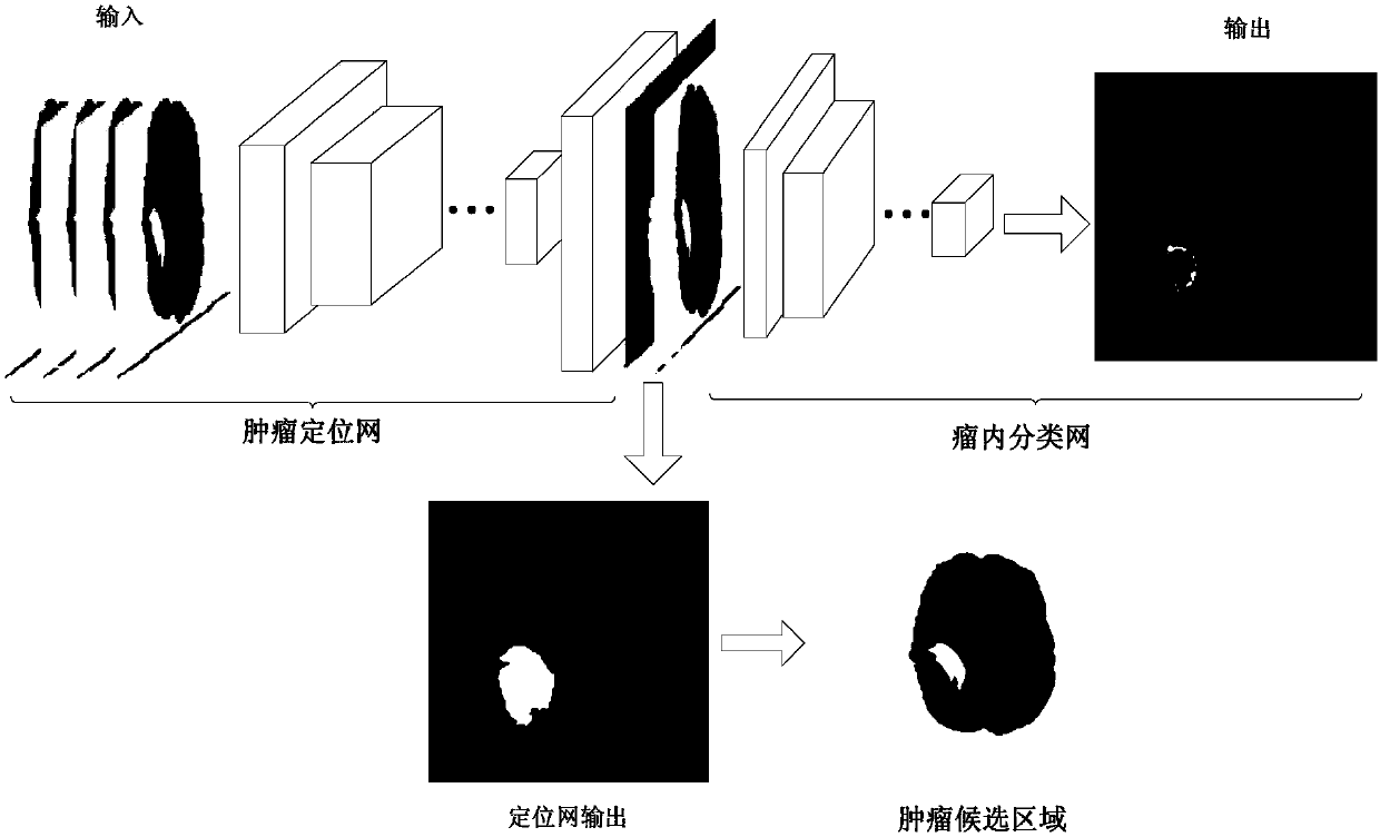
[0006] In the existing MRI brain tumor segmentation, the image block classification method does not use global context features and the segmentation speed is slow, while the training samples of the fully convolutional network pixel-level classification method are seriously unbalanced, resulting in inaccurate segmentation boundaries of small regions. The present invention provides a An MRI brain tumor localization and intratumoral segmentation method based on a deep cascaded convolutional network. This method divides the segmentation process into two stages: complete tumor area localization and intratumoral sub-area segmentation, by building a deep cascaded hybrid convolutional neural network. Fast and accurate localization and intratumoral subregion segmentation of multimodal MRI brain tumors

Method used


Image

  • MRI (Magnetic Resonance Imaging) brain tumor localization and intratumoral segmentation method based on deep cascaded convolution network
  • MRI (Magnetic Resonance Imaging) brain tumor localization and intratumoral segmentation method based on deep cascaded convolution network
  • MRI (Magnetic Resonance Imaging) brain tumor localization and intratumoral segmentation method based on deep cascaded convolution network

Examples


Embodiment Construction

[0067] In order to make the technical means, creative features, goals and effects achieved by the present invention easy to understand, the present invention will be further described below in conjunction with specific illustrations.

[0068] In describing the present invention, it is to be understood that the terms "longitudinal", "radial", "length", "width", "thickness", "upper", "lower", "front", "rear", The orientation or positional relationship indicated by "left", "right", "vertical", "horizontal", "top", "bottom", "inner", "outer", etc. is based on the orientation or positional relationship shown in the drawings , is only for the convenience of describing the present invention and simplifying the description, but does not indicate or imply that the referred device or element must have a specific orientation, be constructed and operated in a specific orientation, and thus should not be construed as limiting the present invention. In the description of the present inventi...


PUM


Abstract

The invention provides an MRI (Magnetic Resonance Imaging) brain tumor localization and intratumoral segmentation method based on a deep cascaded convolution network, which comprises the steps of building a deep cascaded convolution network segmentation model; performing model training and parameter optimization; and carrying out fast localization and intratumoral segmentation on a multi-modal MRIbrain tumor. According to the MRI brain tumor localization and intratumoral segmentation method provided by the invention based on the deep cascaded convolution network, a deep cascaded hybrid neuralnetwork formed by a full convolution neural network and a classified convolution neural network is constructed, the segmentation process is divided into a complete tumor region localization phase andan intratumoral sub-region localization phase, and hierarchical MRI brain tumor fast and accurate localization and intratumoral sub-region segmentation are realized. Firstly, the complete tumor region is localized from an MRI image by adopting a full convolution network method, and then the complete tumor is further divided into an edema region, a non-enhanced tumor region, an enhanced tumor region and a necrosis region by adopting an image classification method, and accurate localization for the multi-modal MRI brain tumor and fast and accurate segmentation for the intratumoral sub-regions are realized.

Description

technical field [0001] The invention relates to the technical field of medical image analysis, in particular to an MRI brain tumor localization and intratumoral segmentation method based on a deep cascaded convolutional network. Background technique [0002] Brain tumor is a major disease that seriously endangers human health. Among them, glioma is the main type of malignant brain tumor. Although it is uncommon, it has a very high fatality rate. According to literature statistics, the average survival time of high-grade glioma is 14 months. Magnetic Resonance Imaging (MRI) is the most commonly used method for the examination and diagnosis of brain tumors in clinical practice. Accurately segmenting brain tumors and intratumoral structures from MRI images is of great value for neuropathological analysis and accurate diagnosis. Provide important support for surgical planning, radiotherapy and chemotherapy plan formulation and prognosis assessment. [0003] The segmentation o...

Claims


Application Information

Patent Timeline
Patent Type & Authority Applications(China)
IPC IPC(8): G06T7/00G06T7/11G06N3/04G06N3/08
CPCG06N3/084G06T7/0012G06T7/11G06T2207/30096G06T2207/30016G06T2207/20081G06T2207/20084G06T2207/20021G06T2207/10088G06N3/045
Inventor 崔少国张建勋
Owner CHONGQING NORMAL UNIVERSITY
Learn More