
Method of improving PET (positron emission tomography) image reconstruction quality by constructing virtual DOI (depth of interaction) and corresponding system matrix

A system matrix and image reconstruction technology, applied in the field of nuclear medicine PET image reconstruction, can solve the problems of low resolution, meaningless, and reduced image spatial resolution of PET images, and achieve the effect of improving image quality and wide application.

Inactive Publication Date: 2018-08-21
WUHAN UNIV
View PDF6 Cites 5 Cited by
  • Summary
  • Abstract
  • Description
  • Claims
  • Application Information

AI Technical Summary

Problems solved by technology

This method can greatly improve the counting efficiency of the detector, and the calculation is simple and convenient, but at the same time, it also ignores the position depth of the photon when it is detected in the scintillation crystal, which will cause a large error in the calculation of the photon emission position, resulting in image distortion. Spatial resolution drops
The low resolution of PET images is also a major criticism in the development of PET technology.
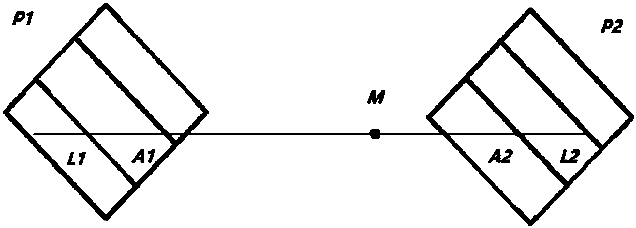
like figure 1 Shown: The traditional PET imaging equipment summarizes a certain LOR event generated by the source A into a pair of detectors P 1 P 2 Generated at a point on the center line of , the calculated source is at P 1 P 2 A certain point on the line, but in fact, the source of the LOR event is at point A, so the tumor position will be deviated after image reconstruction
[0004] In order to solve this problem, a detector that can detect the DOI (depth of interaction) of photons in the crystal can be redesigned and manufactured on the physical hardware, but the physical method has several fatal shortcomings: one is the current detector The manufacturing process is difficult, too thin to capture photons, too thick and meaningless; second, it will make the detector more complicated and lead to higher costs; third, many existing PET devices do not have the conditions to upgrade the hardware, not only need to re-match the PET series Other modules in the system also need to retest the system performance

Method used


Image

  • Method of improving PET (positron emission tomography) image reconstruction quality by constructing virtual DOI (depth of interaction) and corresponding system matrix
  • Method of improving PET (positron emission tomography) image reconstruction quality by constructing virtual DOI (depth of interaction) and corresponding system matrix
  • Method of improving PET (positron emission tomography) image reconstruction quality by constructing virtual DOI (depth of interaction) and corresponding system matrix

Examples


Embodiment Construction

[0025] The content of the present invention will be further introduced below in conjunction with the drawings. The specific implementation steps of the embodiments of the present invention are as follows:

[0026] Step 1. Divide the virtual DOI: divide the scintillation crystal in the PET detector ring into several areas corresponding to different depths of action (DOI) to increase the geometric range of the detector.

[0027] Unlike physical detectors that can detect actual DOI, these areas are virtual. According to the angle of the detector and the geometric range of coverage, the number of virtual DOI can be adjusted freely, and the number of divisions can be determined by the geometric distribution of the PET detector and the size of the crystal. For example, the geometric range covered by the detector itself is very small, and a few virtual DOI can be divided. For the edge part of the crystal (which requires more accurate detection of gamma photons in a larger range), a large...


PUM


Abstract

The invention discloses a method of improving PET (positron emission tomography) image reconstruction quality by constructing a virtual DOI (depth of interaction) and a corresponding system matrix. The method comprises the following steps: 1) virtual DOIs are divided: a scintillation crystal in a PET detector ring is divided into a plurality of regions which correspond to different DOIs; 2) the detection probability of a virtual DOI is calculated: the virtually-divided DOI regions are paired, and the probability of each pair being detected at a different signal source position is calculated; 3) a virtual LOR (Line of response) event in the virtual DOI is divided: one LOR event in one original pair of crystals is divided to multiple sub events in the virtual DOI; 4) a conventional system matrix is optimized: multiple factors that affect the image reconstruction quality are considered; 5) image reconstruction is carried out: a statistical iterative algorithm is used to reconstruct an image; and 6) a GPU platform is used for distributed operation, and the data processing time can be greatly reduced. Through the method disclosed in the invention, the resolution of PET-class equipment after image reconstruction can be greatly improved, the location of a tumor can be positioned accurately, and important clinical value and significance are achieved.

Description

Technical field [0001] The invention relates to the field of nuclear medicine PET image reconstruction, in particular to a PET image reconstruction and optimization method based on constructing a virtual DOI and a corresponding system matrix. Background technique [0002] PET (positron emission tomography, positron emission tomography) is an important clinical examination imaging technology in the field of nuclear medicine. The detection principle of PET is to inject a positron emitting nuclide as a specific tracer into the patient's body. The positron emitted by the tracer and the electrons in the human body are annihilated to produce a pair of opposite gamma photons, and this pair of photons is detected The device records and stores it, and the distribution of the tracer in the patient can be obtained through image reconstruction, thereby realizing early detection and positioning of the tumor. [0003] The detection device in the PET equipment is generally composed of a similar ...

Claims


Application Information

Patent Timeline
IPC IPC(8): G06T11/00
CPCG06T11/003
Inventor 彭浩王成
Owner WUHAN UNIV
Learn More