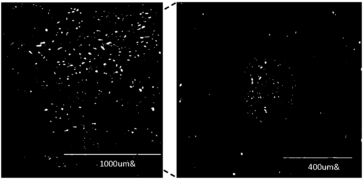
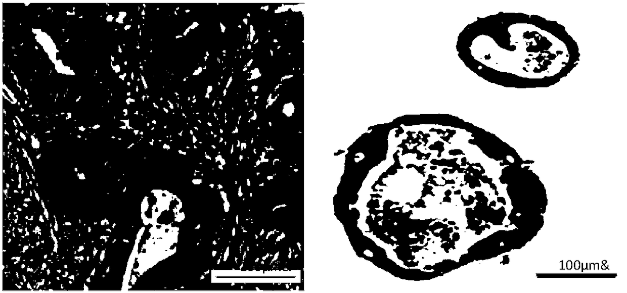
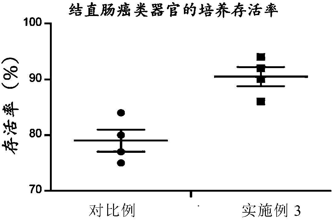
In vitro culture method of colorectal cancer organoid
A colorectal cancer and in vitro culture technology, applied in the field of cell engineering, can solve problems such as low success rate, loss of tumor heterogeneity, limited genetic diversity, etc., achieve high success rate, short time, and improve the effect of culturing rate
- Summary
- Abstract
- Description
- Claims
- Application Information
AI Technical Summary
Problems solved by technology
Method used
Image
Examples
Embodiment 1
[0052] (1) In vitro processing of colorectal cancer biopsy tissue
[0053] Colorectal cancer biopsy tissue was rinsed with 4°C pre-cooled PBS until the supernatant was clear.
[0054] Use a disposable scalpel to cut the tissue into small pieces in a 6cm or 10cm cell culture dish, and the size of the tissue piece is about 5mm.
[0055] Use 20ml of enzymatic digestion solution to transfer the tissue pieces to 50ml Falcon centrifuge tubes. The enzyme digestion solution is composed of: DMEM+2% FBS, 70 units / ml collagen hydrolase, 120 μg / ml neutral protease, 2900 units / ml deoxyribonuclease, 1X penicillin, streptomycin mixed solution.
[0056] Seal the centrifuge tube and incubate it at 37°C for 50 minutes, using a shaker to gently mix the sample during the incubation.
[0057] After the incubation, use a pipette to break up the aggregated tissue clumps, and collect the cells filtered with a 70 μM filter membrane, that is, colorectal cancer single cells.
[0058] Colorectal cance...
Embodiment 2
[0067] (1) In vitro processing of colorectal cancer biopsy tissue
[0068] Colorectal cancer biopsy tissue was rinsed with 4°C pre-cooled PBS until the supernatant was clear.
[0069] Use a disposable scalpel to cut the tissue into small pieces in a 6cm or 10cm cell culture dish, and the size of the tissue piece is about 5mm.
[0070] Use 20ml of enzymatic digestion solution to transfer the tissue pieces to 50ml Falcon centrifuge tubes. The enzyme digestion solution is composed of: DMEM+3% FBS, 80 units / ml collagen hydrolase, 130 μg / ml neutral protease, 3100 units / ml deoxyribonuclease, 1X penicillin, streptomycin mixed solution.
[0071] Seal the centrifuge tube and incubate it at 37°C for 70 minutes, using a shaker to mix the sample gently during the incubation.
[0072] After the incubation, use a pipette to break up the aggregated tissue clumps, and collect the cells filtered with a 70 μM filter membrane, that is, colorectal cancer single cells.
[0073] Colorectal cance...
Embodiment 3
[0081] (1) In vitro processing of colorectal cancer biopsy tissue
[0082] Colorectal cancer biopsy tissue was rinsed with 4°C pre-cooled PBS until the supernatant was clear.
[0083] Use a disposable scalpel to cut the tissue into small pieces in a 6cm or 10cm cell culture dish, and the size of the tissue piece is about 5mm.
[0084] Use 20ml of enzymatic digestion solution to transfer the tissue pieces to 50ml Falcon centrifuge tubes. The enzyme digestion solution is composed of: DMEM+2.5% FBS, 75 units / ml collagen hydrolase, 125 μg / ml neutral protease, 3000 units / ml deoxyribonuclease, 1X penicillin, streptomycin mixed solution.
[0085] Seal the centrifuge tube and incubate it at 37°C for 60 minutes, using a shaker to mix the sample gently during the incubation.
[0086] After the incubation, use a pipette to break up the aggregated tissue clumps, and collect the cells filtered with a 70 μM filter membrane, that is, colorectal cancer single cells.
[0087]Colorectal canc...
PUM
 Abstract
Description
Claims
Application Information
 - R&D
- Intellectual Property
- Life Sciences
- Materials
- Tech Scout
- Unparalleled Data Quality
- Higher Quality Content
- 60% Fewer Hallucinations
Browse by: Latest US Patents, China's latest patents, Technical Efficacy Thesaurus, Application Domain, Technology Topic, Popular Technical Reports.
© 2025 PatSnap. All rights reserved.Legal|Privacy policy|Modern Slavery Act Transparency Statement|Sitemap|About US| Contact US: help@patsnap.com


