
Stable preservation diluent, antinuclear antibody detection reagent and preparation method and application thereof

An anti-nuclear antibody and detection reagent technology, applied in the field of anti-nuclear antibody detection kits, can solve the problems of temperature sensitivity, complex three-dimensional structure, difficult standardization and automation, etc.

Active Publication Date: 2020-10-20
SHENZHEN NEW INDS BIOMEDICAL ENG
View PDF4 Cites 0 Cited by
  • Summary
  • Abstract
  • Description
  • Claims
  • Application Information

AI Technical Summary

Problems solved by technology

Indirect immunofluorescence is still the gold standard for ANA screening. Generally, mouse hepatocytes or Hep-2 cell slices are fixed on glass slides as the target antigen. Positive, the detection time is long, the result is difficult to judge, and it is difficult to standardize and automate; enzyme-linked immunosorbent assay, western blot and dot immunocombination methods have become more and more widely used in ANA detection in recent years. Naturally recombined and purified antigens are coated on microwell plates or fiber membranes. This method can avoid various cytoplasmic interferences to a certain extent. Compared with indirect immunofluorescence, it has better specificity, but it also has cumbersome operations, accuracy and precision. Poor accuracy, limited detection item portfolio, difficulty in automation and technical bottlenecks such as high-throughput quantitative detection
[0006] The target antigens of antinuclear antibodies are mainly extracted from Hep-2 cells, HeLa cells, pig heart tissue or calf thymus tissue, or obtained through prokaryotic cell expression or eukaryotic cell expression, but the target antigens in the cytoplasm and nucleus are complex due to the three-dimensional structure Most of them are complex proteins, and some of the ribonucleoprotein complexes need to be connected by RNA, which makes the target antigen sensitive to temperature after extraction and purification, and sensitive to RNase, DNase and protease
At present, ANA target antigens need to be stored at -20°C or even -70°C, which is not conducive to the development of high-sensitivity quantitative diagnostic reagents

Method used


Image

  • Stable preservation diluent, antinuclear antibody detection reagent and preparation method and application thereof
  • Stable preservation diluent, antinuclear antibody detection reagent and preparation method and application thereof
  • Stable preservation diluent, antinuclear antibody detection reagent and preparation method and application thereof

Examples


preparation example Construction

[0089] The preparation method of the above-mentioned antinuclear antibody detection reagent of one embodiment, comprises the steps:

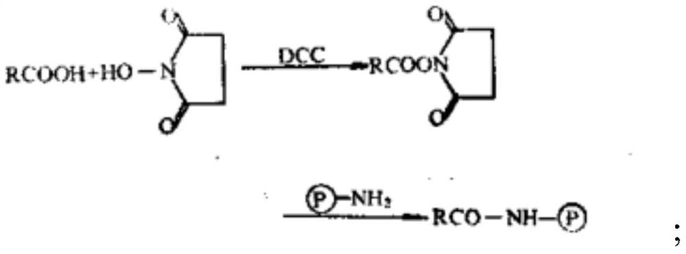
[0090] S10, mixing magnetic microspheres, N-hydroxysuccinimide solution and cross-linking agent solution to obtain activated magnetic microspheres, mixing activated magnetic microspheres with anti-nuclear antibody target antigen to obtain anti-nuclear antibody target antigen and Couplings for magnetic microspheres.

[0091] Antinuclear antibody target antigens can be natural antigens, recombinant antigens, synthetic peptides, HepG2 cell lysis antigens, Hep2 cell lysis antigens, and Hela cell lysis antigens.

[0092] Preferably, the magnetic microspheres need to be cleaned in advance, and can be washed 3 to 5 times with the first washing buffer.

[0093] The first washing buffer can be phosphate buffer or carbonate buffer.

[0094] Magnetic microspheres with -COOH, -NH 2 Or -OH magnetic microspheres, the particle size of the magnetic microsphe...

Embodiment 1

[0129] 1. Take 15 mg of magnetic microspheres, wash them with PBS buffer 3 times, add 0.15 mL of EDC solution with a concentration of 10 mg / mL and 0.15 mL of NHS solution with a concentration of 10 mg / mL for 2 hours, wash 3 times, and then add 0.2 mg The histone antigen was shaken and coated for 2 hours at room temperature to obtain a coupled product of the histone antigen and magnetic microspheres.

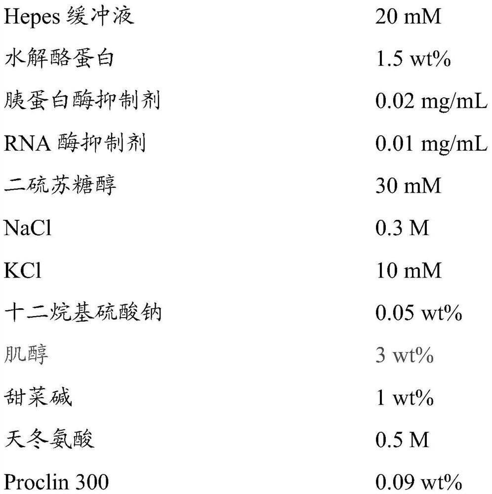
[0130] 2. Prepare stable storage diluent according to the following components and concentrations:

[0131]

[0132] Finally, the pH was adjusted to 6.5 with HCl solution.

[0133] 3. Suspend the above-mentioned coupled substance of histone antigen and magnetic microspheres in the above-mentioned stable storage diluent, and prepare a 1 mg / mL suspension to obtain an antinuclear antibody detection reagent.

Embodiment 2

[0135] 1. Take 15 mg of magnetic microspheres, wash them with PBS buffer 3 times, add 0.15 mL of DCC solution with a concentration of 10 mg / mL and 0.15 mL of NHS solution with a concentration of 10 mg / mL for 2 hours, wash 3 times, and then add 0.2 mg The histone antigen was shaken and coated for 2 hours at room temperature to obtain a coupled product of the histone antigen and magnetic microspheres.

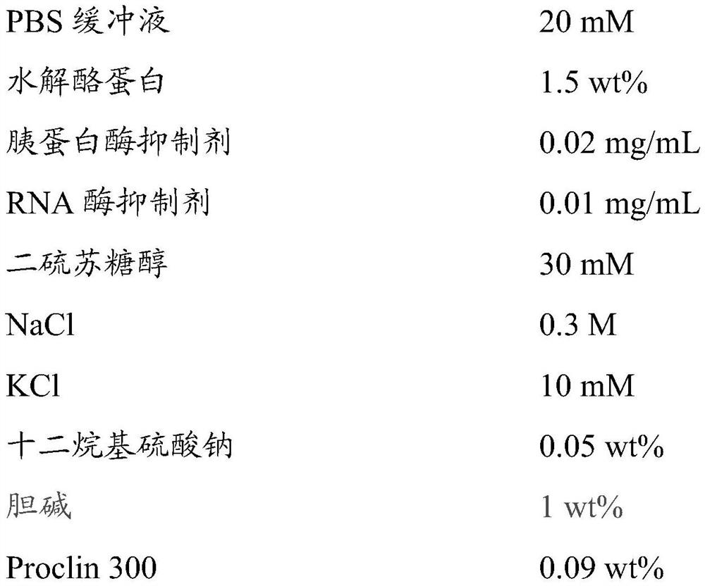
[0136] 2. Prepare stable storage diluent according to the following components and concentrations:

[0137]

[0138] Finally, the pH was adjusted to 6.5 with HCl solution.

[0139] 3. Suspend the above-mentioned coupled substance of histone antigen and magnetic microspheres in the above-mentioned stable storage diluent, and prepare a 1 mg / mL suspension to obtain an antinuclear antibody detection reagent.


PUM

PropertyMeasurementUnit
particle diameteraaaaaaaaaa

Abstract

The invention discloses stable preservation diluent, anti-nuclear antibody detection reagent for the stable preservation diluent, a preparation method thereof and an anti-nuclear antibody detection kit of the anti-nuclear antibody detection reagent. The pH of the stable preservation diluent is 5-8, and the stable preservation diluent includes a buffer solution, salt, osmotic substances and a preservative; the osmotic substances are selected from at least one substance of polyol, methylamine and amino acid. The anti-nuclear antibody detection reagent suspends a coupling matter of anti-nuclear antibody target antigens and magnetic microspheres in the stable preservation diluent for storage to stably store the diluent including the buffer solution, the salt, the osmotic substances and the preservative, thereby providing the coupling matter of the anti-nuclear antibody target antigens and the magnetic microspheres with a stable liquid storage environment.

Description

technical field [0001] The present invention relates to the field of immune detection, in particular to a stable storage diluent, an anti-nuclear antibody detection reagent comprising the stable storage diluent and a preparation method thereof, and an anti-nuclear antibody detection kit comprising the above-mentioned anti-nuclear antibody detection reagent. Background technique [0002] Autoimmune diseases are a type of diseases in which the body produces pathological immune responses against self-antigens due to immune dysfunction, causing organ or system damage. At present, the pathogenesis of autoimmune diseases is still not fully understood. It is generally believed that autoimmune diseases are caused by genetically susceptible individuals under the joint action of environmental factors such as infection, ultraviolet rays, tumors, and drugs. [0003] Antinuclear antibody (antinuclear antibody, ANA) is a general term for a group of autoantibodies that use various compone...

Claims


Application Information

Patent Timeline
Patent Type & Authority Patents(China)
IPC IPC(8): G01N33/543G01N33/531
CPCG01N33/531G01N33/54326
Inventor 饶微张云袁锦云方中刚李婷华程然
Owner SHENZHEN NEW INDS BIOMEDICAL ENG
Learn More