
Magnetic bead for treatment under endoscope

A magnetic bead and magnet technology, applied in the field of medical devices, can solve the problems of unsatisfactory peeling surface or visual field exposure, uneasy operation, low tension, etc., and achieve the effect of facilitating operation, improving safety and clear vision

Inactive Publication Date: 2018-05-22
WEST CHINA HOSPITAL SICHUAN UNIV
View PDF8 Cites 3 Cited by
  • Summary
  • Abstract
  • Description
  • Claims
  • Application Information

AI Technical Summary

Problems solved by technology

After endoscopic local injection, the lifting degree of the submucosa may not be able to achieve the ideal effect, resulting in low tension, peeling surface or visual field exposure is not ideal
Clinically, there are methods such as using dental floss to hang the thread to increase the tension, but the operation is not simple enough, and the efficiency needs to be further improved

Method used


Image

  • Magnetic bead for treatment under endoscope
  • Magnetic bead for treatment under endoscope
  • Magnetic bead for treatment under endoscope

Examples


Embodiment 1

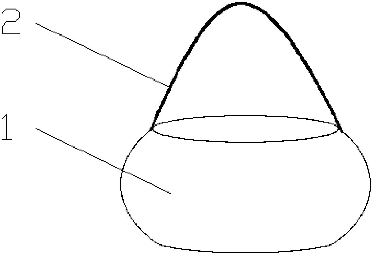
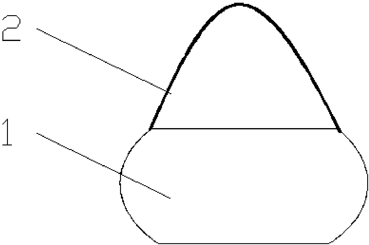
[0034] Such as figure 1 , 2 As shown, in this embodiment, the magnetic poles of the magnet bead 1 are at its upper and lower ends, and the two ends of the fixing wire 2 are fixedly connected to the upper end of the magnet bead 1. The end surfaces of the upper and lower ends of the magnet beads 1 are planes parallel to each other. The cross section of the magnet beads 1 is circular. The diameter of the cross section of the magnet bead 1 is greater than the height of the magnet bead 1. Preferably, the magnet beads 1 have a drum shape. The magnet beads 1 have a solid structure.

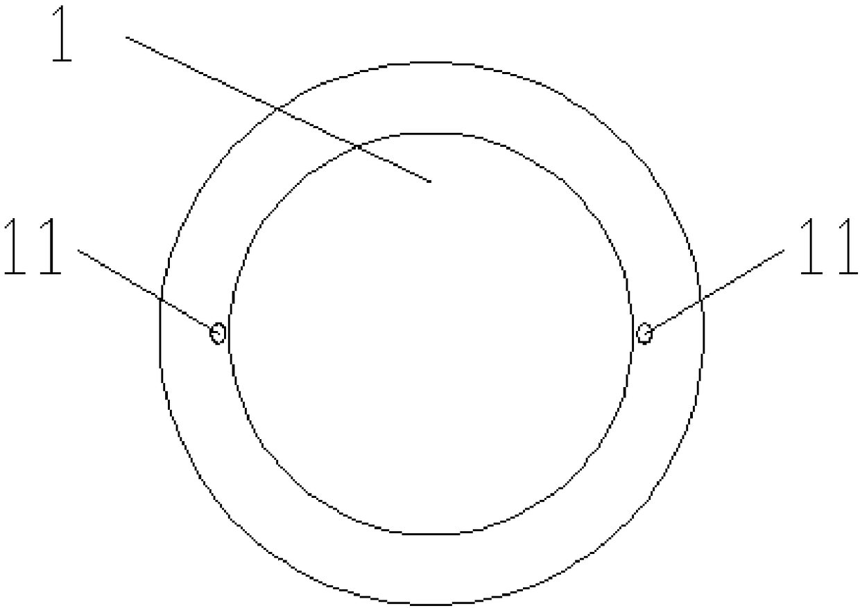
[0035] Such as image 3 As shown, two opposite sides of the magnet bead 1 are respectively provided with a fixed line connection port 11, and both ends of the fixed line 2 are respectively buried in one of the fixed line connection ports 11, so that the fixed line 2 is tightly connected to the magnet bead 1. side.

[0036] The distance between the two fixing wire connection ports 11 is 0.5 cm-1 cm. The l...

Embodiment 2

[0039] The difference between this embodiment and embodiment 1 lies in: Figure 5 , 6 As shown in 7, the magnet bead 1 is a ferromagnetic sphere, and the two opposite sides of the magnet bead 1 are respectively provided with a fixed line connection port 11. Preferably, the fixed line connection port 11 is provided in the middle position of the magnet bead 1, and the fixed line 2 The two ends of the cable are respectively buried in one of the fixed line connection ports 11.

Embodiment 3

[0041] The difference between this embodiment and embodiment 2 lies in: Figure 8 , 9 , 10, the left and right sides of the magnet beads 1 are flat surfaces, and the fixed line connection port 11 is provided in the center of the flat surface.


PUM

PropertyMeasurementUnit
Lengthaaaaaaaaaa
Diameteraaaaaaaaaa
Lengthaaaaaaaaaa

Abstract

The invention relates to a magnetic bead for treatment under an endoscope. The magnetic bead comprises at least one magnet bead and a fixing line. The fixing line is arranged on the at least one magnet bead. The two ends of the fixing line are fixedly connected with the magnet bead; the magnet bead is a ferromagnetic bead body or the upper and lower end faces of the magnet bead are parallel planes. The two opposite sides on the magnet bead are each provided with a fixing line connecting opening, and the two ends of the fixing line are embedded into one fixing line connecting opening. When multiple magnet beads are included, the multiple magnet bead bodies are connected in series to form a magnet bead string. The single magnet bead body or the magnet bead string can provide traction for a surgical target portion where the stripping face is not well exposed, the operation space is narrow, and the view is difficult, tension is generated, the surgical view is clearer, operation is facilitated, and the safety is improved.

Description

Technical field [0001] The present invention relates to the technical field of medical devices, in particular to a magnetic bead for endoscopic treatment. Background technique [0002] With the development of digestive endoscopy diagnosis and treatment technology, the diagnosis rate of early gastrointestinal tumors continues to increase, the number of cases of endoscopic mucosal dissection (ESD) has increased significantly, and the requirements for surgical operations continue to increase. After local injection under the endoscope, the lifting degree of the submucosa may not be able to achieve the desired effect, the resulting tension is low, the peeling surface or the exposure of the visual field is not ideal. Clinically, there have been methods such as using dental floss to pull the thread to increase the tension, but the operation is not simple enough, and the efficiency needs to be further improved. Summary of the invention [0003] The present invention aims to provide magne...

Claims


Application Information

Patent Timeline
IPC IPC(8): A61B17/02
CPCA61B17/0218A61B2017/0225
Inventor 胡兵曾宪晖刘伟叶连松张宇航
Owner WEST CHINA HOSPITAL SICHUAN UNIV
Learn More