
Preparation for cell imaging and preparation method and applications thereof

A technology of cell imaging and preparation, which is applied in the field of cell detection, can solve the problems of no practical application value, inconspicuous imaging effect, weak hydrophilicity of the preparation, etc., and achieve a simple preparation method, avoid interference, and good biocompatibility Effect

Inactive Publication Date: 2018-05-04
THE NAT CENT FOR NANOSCI & TECH NCNST OF CHINA
View PDF3 Cites 6 Cited by
  • Summary
  • Abstract
  • Description
  • Claims
  • Application Information

AI Technical Summary

Problems solved by technology

However, there is a problem in the prior art and the cell imaging reported in the above-mentioned documents, that is, when used in vivo, the AIE molecule is not selective for the cells or tissues to be imaged, and thus has no practical value, and, Due to the weak hydrophilicity and strong hydrophobicity of the obtained preparation, in actual use, AIE molecules mostly gather near the cell membrane, and the imaging effect on the inside of the cell is not obvious

Method used


Image

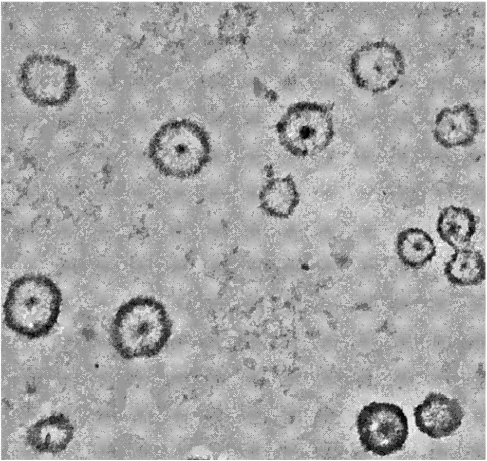
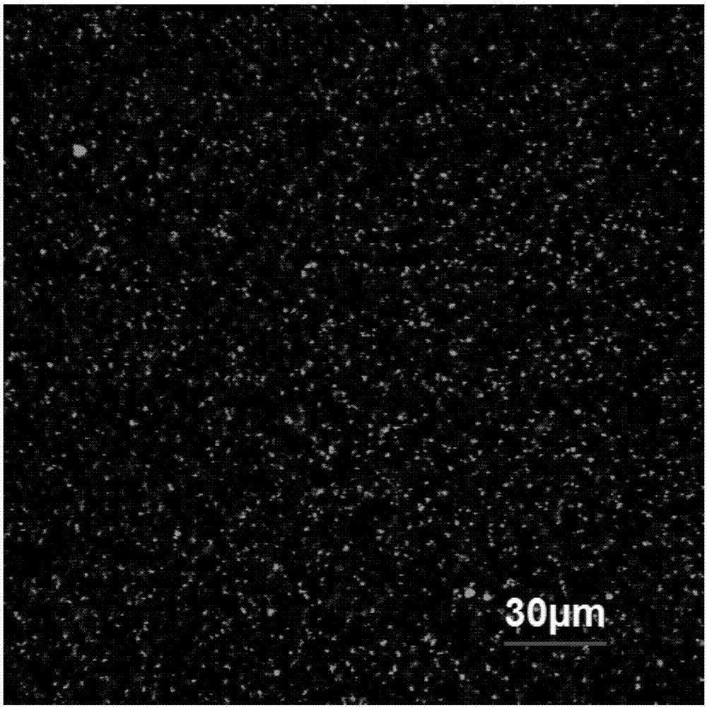
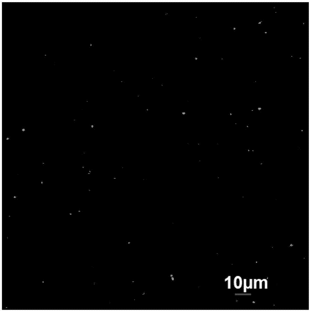
  • Preparation for cell imaging and preparation method and applications thereof
  • Preparation for cell imaging and preparation method and applications thereof
  • Preparation for cell imaging and preparation method and applications thereof

Examples


Embodiment 1

[0049] Formulation 1 was prepared by the following steps:

[0050] Step (1), disperse 5 mg of bispyrene molecules in a solvent obtained by mixing acetone and water at a volume ratio of 1:1, use a magnetic stirrer to mix and stir at a speed of 800 rpm for 20 minutes, and then ultrasonically treat for 40 minutes to make it bispyrene. The pyrene molecules are uniformly dispersed to obtain a bispyrene molecular dispersion with a concentration of 0.3 mg / mL;

[0051] In step (2), take the bispyrene molecular dispersion obtained in step (1), add it to a solution of dimyristoylphosphatidylcholine in acetone in which 550 mg of dimyristoylphosphatidylcholine is dissolved, and ultrasonically treat it for 40 minutes to make it Mix evenly, then put the mixed solution into a rotary evaporator, adjust the rotary evaporator to work at a speed of 50 rpm for 45 minutes, so that all the solvents in it are volatilized and form a uniform and transparent film on the rotating plane of the rotary eva...

Embodiment 2

[0054] Formulation 2 was prepared by the following steps:

[0055] The difference from Example 1 is that the bispyrene molecules used in each step are replaced by MC-4 molecules.

[0056] Example 2 Formulation 2 was obtained.

Embodiment 3

[0058] Formulation 3 was prepared by the following steps:

[0059] The difference from Example 1 is that the dimyristoylphosphatidylcholine used in each step is replaced by phospholipid polyethylene glycol folic acid with a number average molecular weight of 5200.

[0060] Example 3 Formulation 3 was obtained.


PUM

PropertyMeasurementUnit
particle diameteraaaaaaaaaa
concentrationaaaaaaaaaa
pore sizeaaaaaaaaaa

Abstract

The invention relates to a preparation for cell imaging and a preparation method and applications thereof. The preparation comprises phospholipid microspheres with dissolved AIE molecules and a solvent for dispersing phospholipid microspheres. The AIE molecules are fat soluble molecules with an AIE effect. The preparation method of the preparation is very simple. The prepared preparation has goodbiocompatibility and is uniformly dispersed. The particle size is in a range of 100-300 nm and is adjustable. In cells, the structure of phospholipid microspheres is degraded by a surfactant, and thedegradation time is 5-36 hours. Dissolved AIE molecules are released and aggregated. After laser radiation, fluorescence is generated, thus cell imaging and cell labeling are realized; at the same time, two-photon imaging can be realized through long wavelength laser excitation; the images are clear, furthermore, the interference of other fluorescent substances in cytoplasm is avoided, and by using the EPR effect of tumor tissues, the provided preparation can specifically identify and label the tumor cells.

Description

technical field [0001] The invention relates to the field of cell detection, in particular to a preparation for cell imaging, a preparation method and application thereof. Background technique [0002] Liposome (Liposome) is an amphipathic artificial membrane, with phospholipid or phospholipid-like molecules as the main component. Common liposomes in the human body are phospholipid bilayers in cell membranes. Phospholipid or phospholipid-like molecules are in water and aqueous solution, the hydrophilic head is inserted into the water, and the hydrophobic tail extends to the air. After agitation, a spherical liposome structure of bilayer lipid molecules is formed, with a diameter ranging from 25 to 1000 nm. Liposomes have a cell membrane-like structure and good biocompatibility. Some fat-soluble drugs can be encapsulated in liposome bilayers to form microvesicles, which can fuse with cell membranes and deliver drugs into Therefore, liposomes can be used as fat-soluble drug c...

Claims


Application Information

Patent Timeline
Patent Type & Authority Applications(China)
IPC IPC(8): C09K11/06G01N21/64
CPCC09K11/06C09K2211/1011C09K2211/1014C09K2211/1051G01N21/6428G01N2021/6439
Inventor 杨洋李影王磊李峻柏
Owner THE NAT CENT FOR NANOSCI & TECH NCNST OF CHINA
Learn More