
Magnetic resonance imaging rectification method, device and equipment based on 3D shape and appearance measurement

A magnetic resonance imaging and shape measurement technology, applied in the medical field, can solve the problems of prolonged scanning time, single purpose, and inability to measure accurately, and achieve the effects of improving measurement accuracy, improving imaging quality, and speeding up imaging speed

Active Publication Date: 2018-03-27
TSINGHUA UNIV
View PDF9 Cites 2 Cited by
  • Summary
  • Abstract
  • Description
  • Claims
  • Application Information

AI Technical Summary

Problems solved by technology

[0003] At present, a variety of motion artifact correction methods have been proposed at home and abroad. The most important technology is to detect motion by collecting a small amount of magnetic resonance signals (Navigator technology) before scanning. The most advanced navigator can automatically calculate the rigidity. Motion parameter, suitable for head imaging, but it interferes with normal MRI image acquisition, prolongs scan time, and cannot accurately detect non-rigid motion; then it is proposed to develop fast imaging techniques that are not sensitive to motion, including Cartesian and non-Cartesian Parallel imaging of sampling trajectories, Compress Sensing and other technologies, but the image signal-to-noise ratio will be lost, and the self-navigator technology developed by using the characteristics of non-Cartesian sampling trajectories such as radial and propeller will continuously collect k-space centers, using continuously collected As a navigator, the k-space center does not interfere with image acquisition, but it has its own unique reconstruction problems, and the measurement of motion is only to detect the presence or absence, and cannot be accurately measured. There are also many methods to use external equipment to monitor patient motion in MRI scans. Detection and measurement, including the commonly used chest and abdominal belts for measuring respiratory signals, ECG or PPU (Physics Processing Unit, physical computing processor) for measuring heart motion, etc., and field detectors and marker points developed in recent years Optical measurement and other methods, but the chest belt, ECG and PPU are single-purpose, unable to detect patient motion other than physiological signals, and the measurement accuracy of field detectors to detect motion parameters is not high, and the optical measurement method of marker points can only measure non-rigid motion

Method used


Image

  • Magnetic resonance imaging rectification method, device and equipment based on 3D shape and appearance measurement
  • Magnetic resonance imaging rectification method, device and equipment based on 3D shape and appearance measurement
  • Magnetic resonance imaging rectification method, device and equipment based on 3D shape and appearance measurement

Examples


Embodiment Construction

[0033] Embodiments of the present invention are described in detail below, examples of which are shown in the drawings, wherein the same or similar reference numerals designate the same or similar elements or elements having the same or similar functions throughout. The embodiments described below by referring to the figures are exemplary and are intended to explain the present invention and should not be construed as limiting the present invention.

[0034] The following describes the magnetic resonance imaging correction method, device and equipment based on 3D topography measurement according to the embodiments of the present invention with reference to the accompanying drawings. First, the magnetic resonance imaging based on 3D topography measurement according to the embodiments of the present invention will be described with reference to the accompanying drawings corrective method.

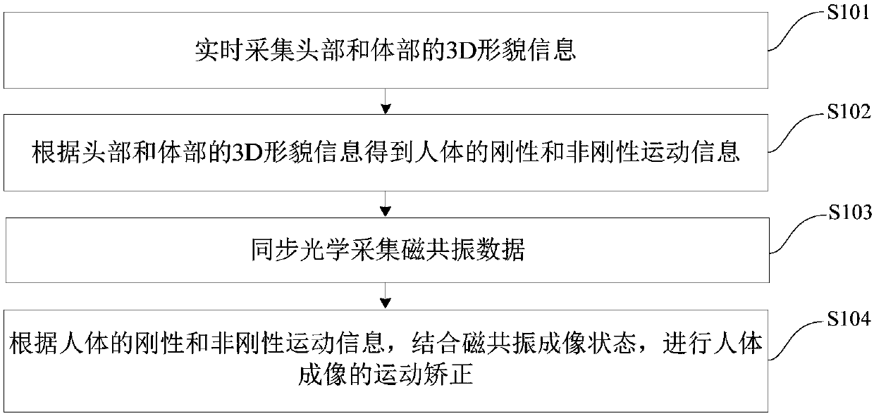
[0035] figure 1 It is a flow chart of the magnetic resonance imaging correction method b...


PUM


Abstract

The invention discloses a magnetic resonance imaging rectification method, device and equipment based on 3D shape and appearance measurement, and the method comprises the steps: collecting the shape and appearance information of a head part and a body part; obtaining the rigid and non-rigid movement information of a human body according to the shape and appearance information of the head part andthe body part; synchronously collecting the magnetic resonance data; and carrying out the movement rectification of human body imaging according to the rigid and non-rigid movement information of thehuman body through combining with the magnetic resonance imaging state. The method can achieve the precise measurement of the rigid and non-rigid movement of the human body according to the shape andappearance information of the head part and the body part, so as to achieve the movement rectification of human body imaging. The method is simpler in operation, can effectively speed up the imaging,improves the imaging quality, and improves the measurement precision.

Description

technical field [0001] The invention relates to the field of medical technology, in particular to a magnetic resonance imaging correction method, device and equipment based on 3D shape measurement. Background technique [0002] Due to its advantages of high spatial resolution and high contrast of soft tissue imaging, magnetic resonance imaging is currently an important clinical imaging diagnostic tool and plays an indispensable role in the diagnosis of major diseases such as cancer, liver disease, and cardiovascular and cerebrovascular diseases. However, limited by the physical principle of its imaging, the imaging speed of magnetic resonance is relatively slow. In actual clinical use scenarios, it is difficult for the patient to keep still during the long-term imaging process, and the movement of the patient will affect the imaging quality. Low and artifacts can even interfere with doctor's diagnosis, and in the worst case can lead to misdiagnosis. For imaging, the patient...

Claims


Application Information

Patent Timeline
IPC IPC(8): G01R33/565
CPCG01R33/56509
Inventor 陈慧军齐海坤章强
Owner TSINGHUA UNIV
Learn More