
Diagnostic imaging system comprising a device for facilitating breast examinations

A breast and irradiation system technology, applied in the directions of diagnosis, sonic diagnosis, infrasound diagnosis, etc., can solve the problems of patient shape adaptation, inconvenience, inconvenience, etc.

Inactive Publication Date: 2018-02-06
NOVAURA SRL
View PDF14 Cites 2 Cited by
  • Summary
  • Abstract
  • Description
  • Claims
  • Application Information

AI Technical Summary

Problems solved by technology

[0009] However, this ductoscopy system exhibits significant drawbacks, mainly related to the overall size of the device
[0010] Furthermore, the device is structurally complex and, therefore, difficult to manage
[0011] Another significant drawback of the prior art arises from the inconvenience that the patient must have access to the immobilization area that engages the breast with the support device
Said support device is accommodated in a region of the machine which does not fit morphologically to the patient, whereby the patient must adapt himself in order to properly place the breast on the device, thus resulting in an inconvenient drawback
[0012] Furthermore, the device is particularly inconvenient for an operator who does not have access to the area of ​​the breast that is held fixed to the device (for example, an operator using an ultrasound probe for further control)

Method used


Image

  • Diagnostic imaging system comprising a device for facilitating breast examinations
  • Diagnostic imaging system comprising a device for facilitating breast examinations
  • Diagnostic imaging system comprising a device for facilitating breast examinations

Examples


Embodiment Construction

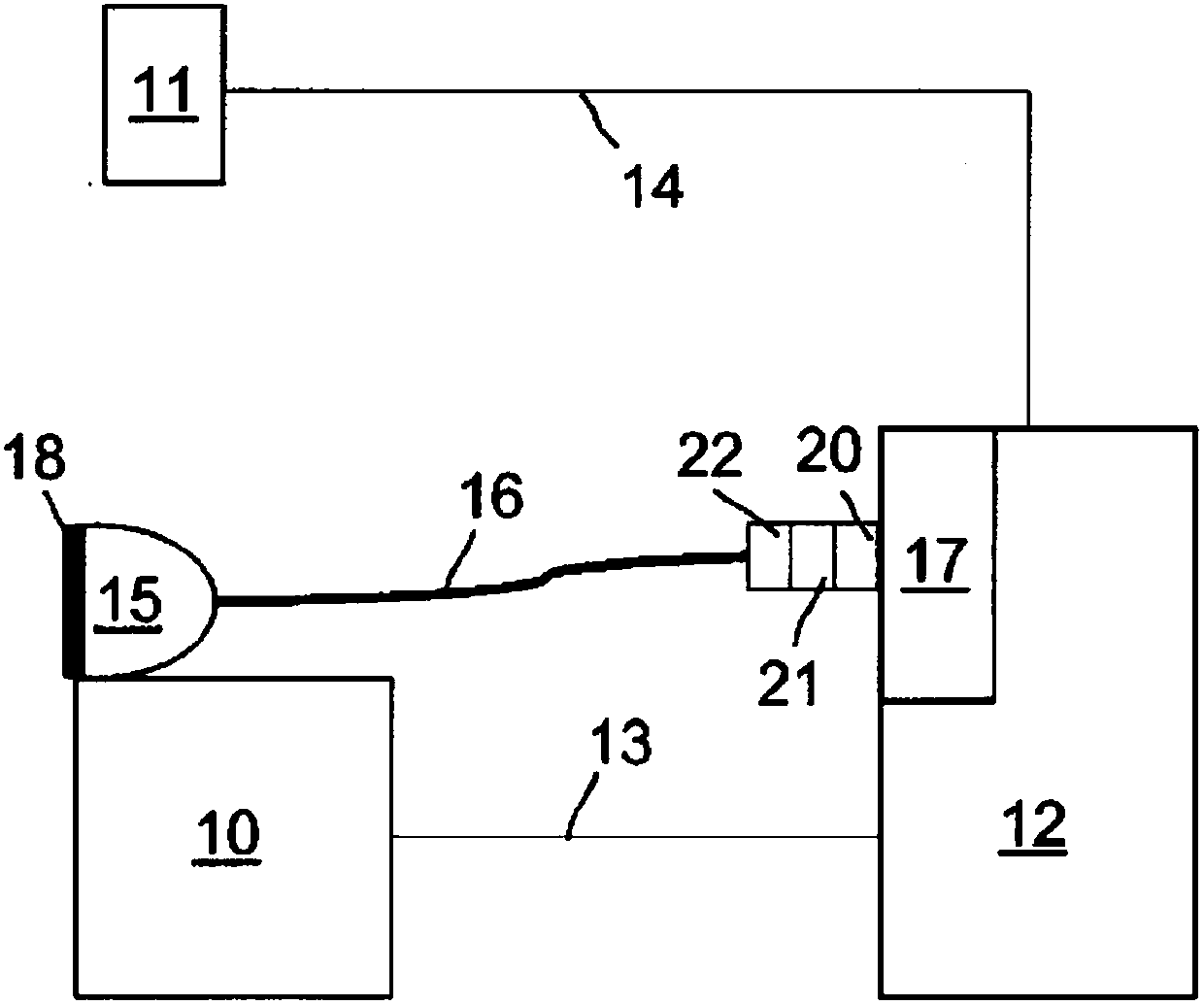
[0030] Referring to the drawings, the diagnostic imaging system according to the present invention includes an illumination system 10 that emits light in the visible light range, for example, 640 nm light, or 750 nm to 1400 nm light in the near infrared light range.

[0031] The camera 11 is opposed to the illumination system 10. The camera 11 is set in a known manner (distance, filter, etc.) so as to acquire an image of the biological tissue to be inspected illuminated by the illumination system 10 below.

[0032] The control center 12 is electrically connected to the illumination system 10 through a cable 13 and is electrically connected to the camera 11 through a cable 14 for controlling, managing and transmitting data.

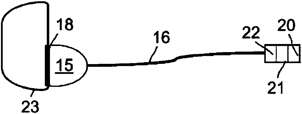
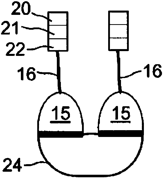
[0033] The breast to be analyzed is placed in a device 15 which is pneumatically connected to a reduced pressure source 17 through a tube 16.

[0034] The device 15 is essentially a thin film made of an elastic material (for example, a silicone type material).

[0...


PUM


Abstract

Diagnostic imaging system and method comprising a device for facilitating breast examinations, comprises: at least one hemispherical portion having a convex outer surface and a concave inner surface defining a containment chamber of a breast; means to wear said hemispherical portion positionable at least around the user's torso to hold the hemispherical portion on said breast; and a connecting tube associated with said hemispherical portion to put in fluid communication the containment chamber with a depressurisation source; the hemispherical portion being made of elastic material to be switchable between a compression condition of the breast and a release condition of the breast.

Description

Technical field [0001] The present invention relates to a diagnostic imaging system including a device to facilitate breast examination. [0002] In particular, the present invention relates to a device for compressing the breast of a patient in an examination for early diagnosis of breast tumors using optical technology. The diagnostic system can also allow the examination to be combined with other diagnostic techniques (for example, ultrasound). Background technique [0003] For women, breast tumors are second only to skin tumors, but the mortality rate is the first. [0004] A particularly disturbing phenomenon is the continuous decline in the average age at which tumors occur, and the insufficient diagnostic capabilities of current screening devices in the case of dense breasts, which are characteristic of women of premenopausal age who are generally in good health. In fact, mammography is not reliable enough for dense breasts. Ultrasound alone may be inefficient if it cannot ...

Claims


Application Information

Patent Timeline
IPC IPC(8): A61B5/00A61B8/00A61B8/08
CPCA61B5/0091A61B5/708A61B8/0825A61B8/085A61B8/40A61B8/4416A61B5/0062A61B8/4245A61B5/00A61B6/502A61B8/403
Inventor 达尼洛·真纳里
Owner NOVAURA SRL
Learn More