
Automatic generation of radiology reports from images and automatic rule out of images without findings

A radiology, automatic technology, applied in the direction of radiological diagnostic instruments, medical reports, medical images, etc., can solve the problem of not providing identification and exclusion

Inactive Publication Date: 2017-11-28
SIEMENS HEALTHCARE GMBH
View PDF4 Cites 7 Cited by
  • Summary
  • Abstract
  • Description
  • Claims
  • Application Information

AI Technical Summary

Problems solved by technology

Conventional systems do not provide any efficient means to identify and exclude multi-dimensional (e.g. 2D / 3D / 4D) images without findings with high confidence

Method used


Image

  • Automatic generation of radiology reports from images and automatic rule out of images without findings
  • Automatic generation of radiology reports from images and automatic rule out of images without findings
  • Automatic generation of radiology reports from images and automatic rule out of images without findings

Examples


Embodiment Construction

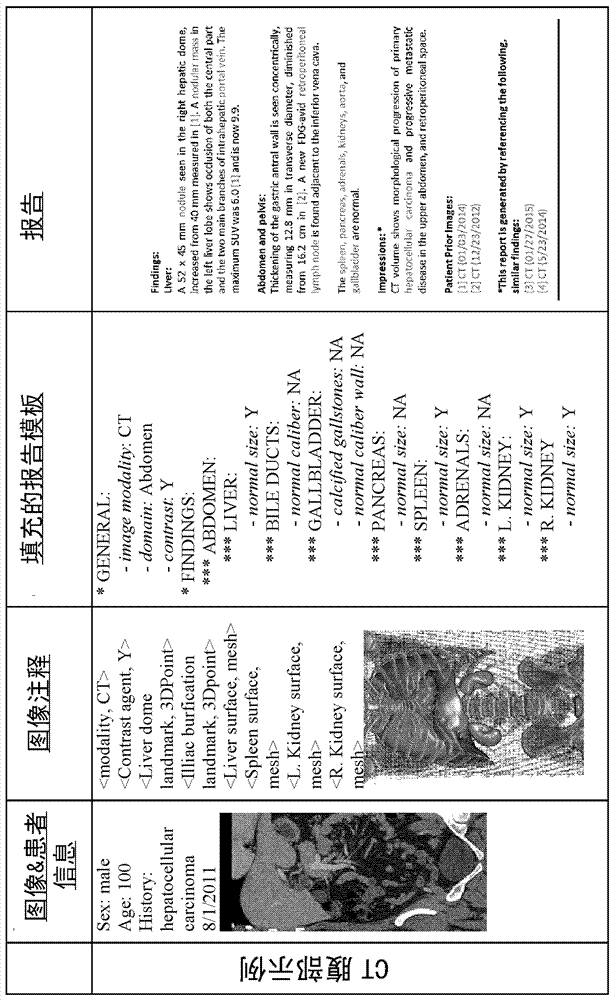
[0025]The following disclosure describes the invention in terms of various embodiments directed to methods, systems, and apparatus for automatically and efficiently parsing medical image data to derive radiological findings. The disclosed techniques can be applied to automatically generate radiology reports by extracting structured report templates and concepts and determining their associated annotations that can be derived from image processing. Additionally, with the ability to eliminate images not found, the disclosed technique can be used to adjust scan acquisitions and filter irrelevant images for screening for diseases such as lung cancer. In addition, the use of standardized fields and templates streamlines comparisons with longitudinal data and similar cases from past reports, which allows this system to rapidly process and interpret current data in the context of historical big data. patient data. The techniques described herein have the potential to not only automa...


PUM


Abstract

A computer-implemented method for automatically generating a radiology report includes a computer receiving an input dataset comprising a plurality of multidimensional patient images and patient information and parsing the input dataset using learned models to determine a clinical domain and relevant image annotations. The computer populates an annotation table using the relevant image annotations and applies one or more domain-specific scriptable rules to populate a report template based on the annotation table. The computer may then generate a natural language radiology report based on the report template.

Description

technical field [0001] The present invention generally relates to methods, systems and apparatus for automatically generating radiology reports from images and automatically excluding images not found. Use of the disclosed methods, systems, and apparatus can be applied to processing information collected from a variety of imaging modalities including, without limitation, computed tomography (CT), magnetic resonance (MR), positron Emission tomography (PET), and ultrasound (US) techniques. Background technique [0002] Current standards of practice for reporting in radiology require clinicians to examine each individual slide in a volume data set (eg MR, CT or PET) and dictate a verbal summary of the findings. This dictation was later transcribed into a free-form written report in which the overall imprint from the images was correlated with non-image patient information (ie age, patient history). This labor-intensive process is subjective and unclear as to which image infor...

Claims


Application Information

Patent Timeline
IPC IPC(8): G06T7/00G06F19/00G06F40/00G16H10/60G16H15/00G16H30/40
CPCG06T7/0012G06T2207/10081G06T2207/10088G06T2207/10104A61B6/4441G06Q10/10G16H40/63G16H10/60G16H50/20G16H15/00A61B6/032A61B6/463A61B6/545G06F40/169G06F40/186G06F40/56G16H30/40G06F40/40
Inventor D.科马尼丘B.乔治斯库刘雯霈周少华
Owner SIEMENS HEALTHCARE GMBH
Learn More