Mesenchymal stem cells tracked by fluorescent and mri dual image functional microspheres and its application
A stem cell, dual imaging technology, applied in cells modified by introducing foreign genetic material, general/multifunctional contrast agents, medical preparations containing active ingredients, etc., can solve problems such as undiscovered molecular probes, and reduce Effects of auto background fluorescence, minimal tissue damage, good biodegradability and biocompatibility
- Summary
- Abstract
- Description
- Claims
- Application Information
AI Technical Summary
Problems solved by technology
Method used
Image

Examples
Embodiment 1
[0036] The preparation method of the fluorescent and MRI double image functional microspheres of the present invention comprises the following steps:
[0037] (1) Weigh 2mg of ring metal iridium compound (Tris(2-(benzo[b]thiophen-2-yl)pyridineiridium(III) or Bis(2-benzo[b]thiophen-2-yl-pyridine)(acetylacetonate) iridium (III)) and 25mg PLA-PEG were mixed and dissolved with 1mL chloroform to obtain an oil phase. Prepare 100 μL of 10mg / mL Gd-DTPA contrast agent as the inner water phase.
[0038](2) The oil phase obtained in step (1) and the internal water phase were ultrasonically mixed 5 times, each time for 2 s (100 W, BILON92-II), to obtain colostrum.
[0039] (3) Add 4mL PVA aqueous solution (5wt%) to the product of step (2), and ultrasonically mix 5 times (2s, 100W) to form a double emulsion.
[0040] (4) Dilute the product of step (3) into PAA aqueous solution (5wt%, 40mL), stir overnight at room temperature, and avoid light to evaporate.
[0041] (5) Use a 50mL high-spe...
Embodiment 2
[0044] The preparation method of MSCs traced by fluorescent and MRI double image functional microspheres of the present invention comprises the following steps:
[0045] (1) HUCMSCs were normally subcultured, and the corresponding specifications were spread on the bottom of ordinary cell culture dishes.
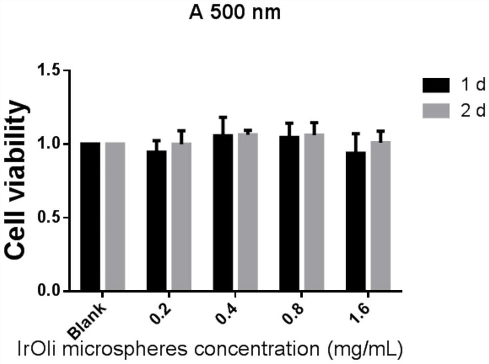
[0046] (2) Mix the microsphere precipitation prepared in Example 1 with the MSCs medium, make a microsphere culture solution (0.2-1mg / mL) and place it in an incubator (37°C, 5% CO 2 ) and incubated for 24h.
[0047] (3) After 24 hours, HUCMSCs adhered to the wall, sucked off the culture medium, added the same volume of microsphere culture medium (0.2-1 mg / mL) incubated in step (2), and placed in an incubator (37 ° C, 5% CO 2 ), continue culturing for 2-4 days, preferably 2 days.
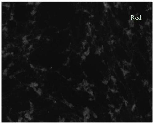
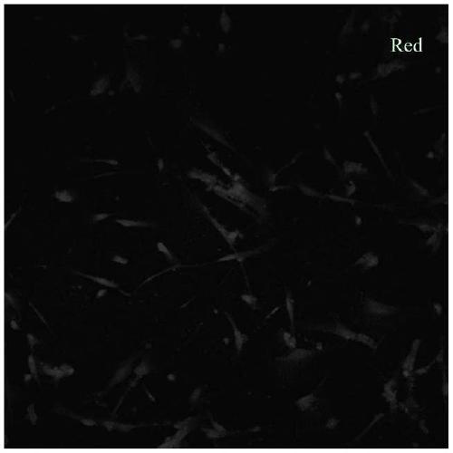
[0048] (4) After 2-4 days, remove the culture medium, wash twice with PBS, immerse in 5% paraformaldehyde for 30 minutes, wash twice with PBS, slide the bottom of the dish to make slide specimens....
Embodiment 3
[0055] The preparation method of the fluorescent and MRI double image functional microspheres of the present invention comprises the following steps:
[0056] (1) Weigh 2 mg of cyclometal iridium compound, mix with 25 mg of PLGA, PLA-PEG, and PLGA-PEG respectively, and dissolve with 1 mL of chloroform respectively to obtain an oil phase. Prepare 100 μL of Gd-DTPA contrast agent as the inner aqueous phase.
[0057] (2) The oil phase obtained in step (1) and the internal water phase were ultrasonically mixed 5 times (2s, 100W, BILON92-II) to obtain a colostrum.
[0058] (3) Add 4mL PVA aqueous solution (5wt%) to the product of step (2), and ultrasonically mix 5 times (2s, 100W) to form a double emulsion.
[0059] (4) Dilute the product of step (3) into PAA aqueous solution (5wt%, 40mL), stir overnight at room temperature, and avoid light to evaporate.
[0060] (5) Use a 50mL high-speed centrifuge tube to collect the solution containing microspheres in step (4), wash it once wi...
PUM
| Property | Measurement | Unit |
|---|---|---|
| particle diameter | aaaaa | aaaaa |
| concentration | aaaaa | aaaaa |
| diameter | aaaaa | aaaaa |
Abstract
Description
Claims
Application Information
 - Generate Ideas
- Intellectual Property
- Life Sciences
- Materials
- Tech Scout
- Unparalleled Data Quality
- Higher Quality Content
- 60% Fewer Hallucinations
Browse by: Latest US Patents, China's latest patents, Technical Efficacy Thesaurus, Application Domain, Technology Topic, Popular Technical Reports.
© 2025 PatSnap. All rights reserved.Legal|Privacy policy|Modern Slavery Act Transparency Statement|Sitemap|About US| Contact US: help@patsnap.com