


Quick Research
Generate reliable direction feasibility study reports for your R&D in just a few steps.

Technical Q&A
Discover and master advanced knowledge NOW. Basics, ideas, possibilities, all at once.

Find Solutions
As an expert in R&D theories, this can generate solutions to your technical problems instantly.

Evaluate Feasibility
Analyze your overall solution with one click, know your potential R&D risks in advance.

Monitor Landscape
Get weekly tech updates, stay abreast of the latest tech innovations and key insights.
Cone-beam CT multi-directional scanning apparatus
A technology of CT scanning and cone beam, which is applied in computerized tomography scanners, instruments for radiological diagnosis, medical science, etc., and can solve problems such as staying in morphological performance, long scanning time, and poor image quality
- Summary
- Abstract
- Description
- Claims
- Application Information
AI Technical Summary
Problems solved by technology
Method used
Image

Examples
Embodiment 1
[0033] Three-dimensional scanning imaging of patients in standing position:
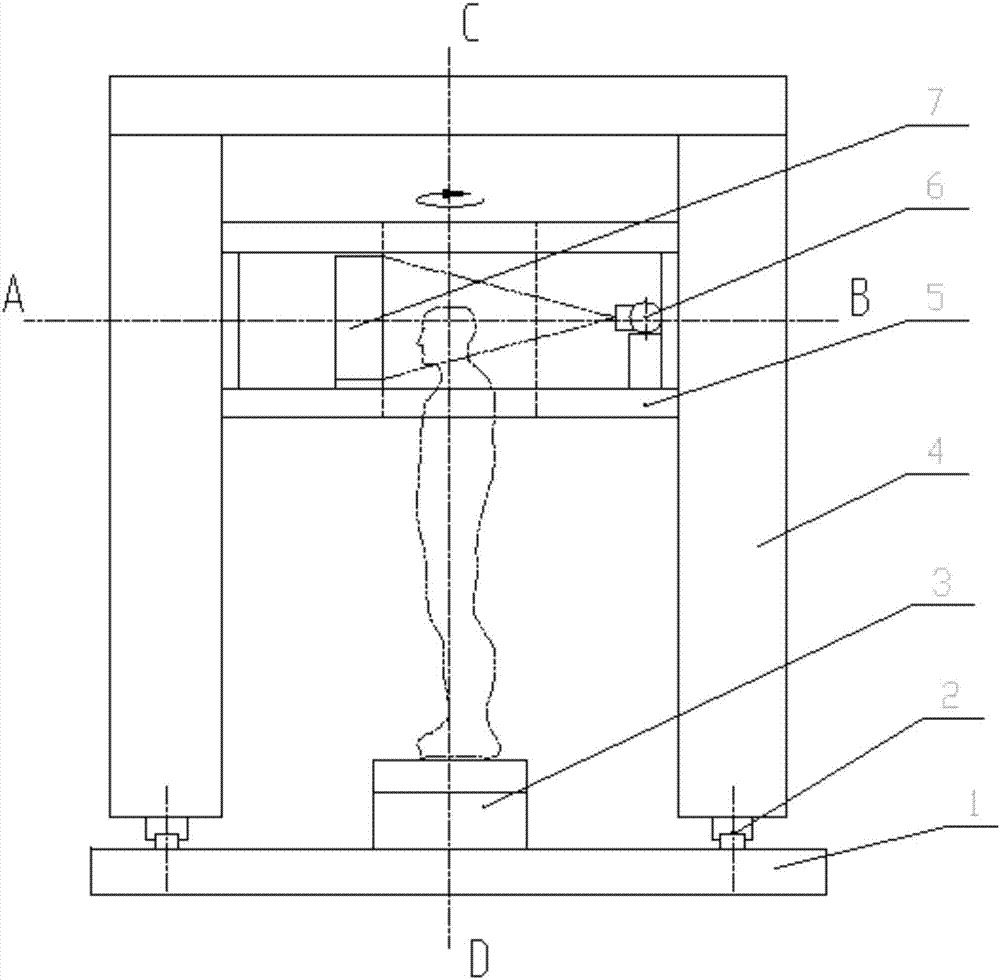
[0034] Such as figure 1 and figure 2As shown, after the operator enters the basic information of the patient, he selects the vertical three-dimensional scanning mode. After the confirmation of the auxiliary system, the patient stands correctly on the standing platform 3. The auxiliary system defines the required scanning inspection range, starts the inspection, and the equipment presses Control the operation of the required logical action: the rotating scanning table 10 rotates, the ball tube 6 emits the beam as required, the dynamic flat panel detector 7 receives the projection information, the control system transmits and saves the data, and after scanning for one week, the scanning frame 5 moves down by one imaging height Carry out the next scan until the defined truncation is scanned, and the machine returns to the initial state. The patient leaves the machine. The data processing system gene...
Embodiment 2
[0036] Three-dimensional scanning imaging of the patient in the supine position:
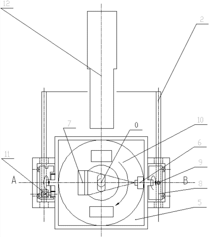
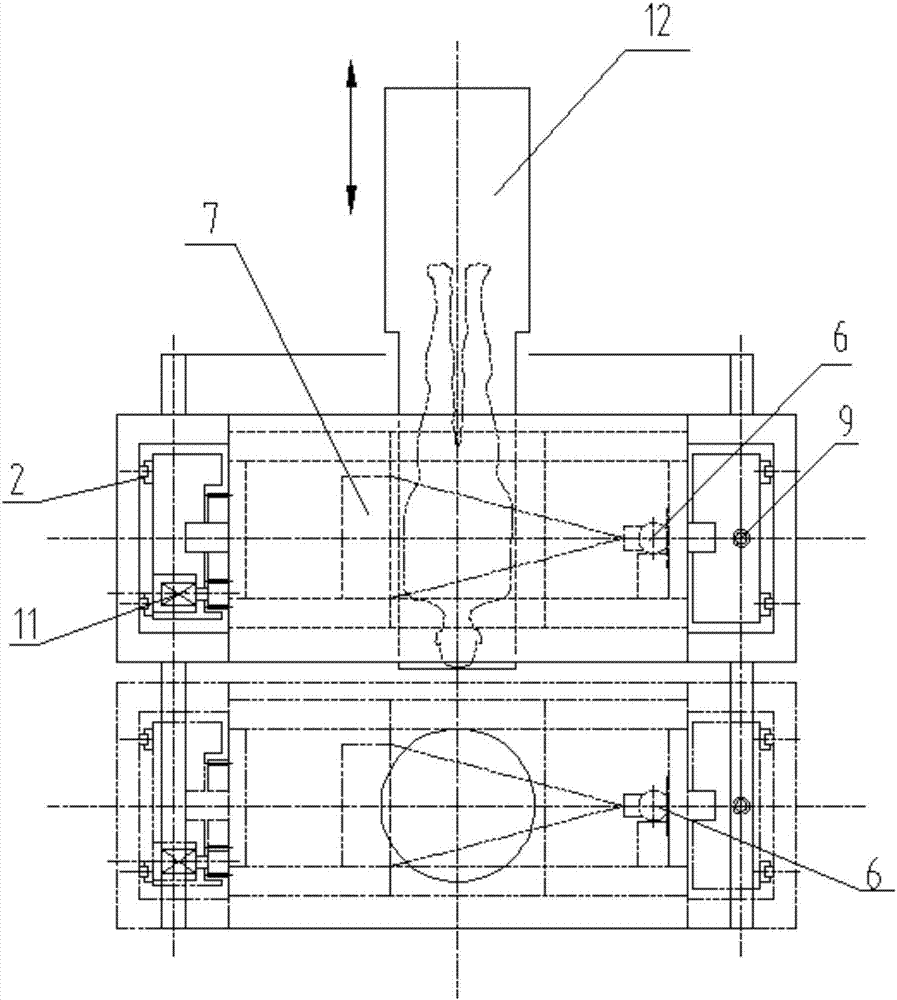
[0037] like image 3 , Figure 4 and Figure 5 As shown, after the operator enters the basic information of the patient, the operator selects the horizontal three-dimensional scanning mode, and the system will switch to the horizontal scanning mode: the scanning frame 5 is turned 90 degrees at the height H Figure 4 state, the scanning couch 12 is lowered to the lowest final initial position.
[0038] Under the confirmation of the auxiliary system, the patient lies correctly on the scanning table 12, defines the required scanning inspection range with the auxiliary system, starts the inspection, and the equipment operates according to the logical action required by the control: the scanning table 12 rises to a predetermined height, moves forward Move to the scanning start position, the rotary scanning table 10 rotates, the ball tube 6 emits the beam as required, the dynamic flat panel detecto...
Embodiment 3
[0040] Patient two-dimensional scan imaging:
[0041] In the scanning process of Embodiment 1 and Embodiment 2, the rotating scanning table 10 is only statically photographed in the front and side positions to complete the set axial imaging range of the human body, and DR imaging in standing and lying states can be realized.
[0042] The cone-beam CT multi-directional scanner of the present invention can change the current situation that it is impossible to obtain three-dimensional bone image data in the upright position of the human body. Three-dimensional cone-beam CT imaging and two-dimensional DR shaping can be performed in the standing and lying positions to obtain three-dimensional images of the human skeletal system and soft tissues with high-quality spatial precision and high-quality density precision. Provide good support for clinical biomechanical assessment, diagnosis, treatment, surgical planning, simulation, etc.
PUM
 Abstract
Description
Claims
Application Information
 - R&D Engineer
- R&D Manager
- IP Professional
- Industry Leading Data Capabilities
- Powerful AI technology
- Patent DNA Extraction
Browse by: Latest US Patents, China's latest patents, Technical Efficacy Thesaurus, Application Domain, Technology Topic, Popular Technical Reports.
© 2024 PatSnap. All rights reserved.Legal|Privacy policy|Modern Slavery Act Transparency Statement|Sitemap|About US| Contact US: help@patsnap.com