Preparation method of human myeloperoxidase immunochromatographic test strip
A myeloperoxidase and immunochromatographic test paper technology, which can be used in analytical materials, measuring devices, instruments, etc., can solve problems such as insufficient accuracy and sensitivity
- Summary
- Abstract
- Description
- Claims
- Application Information
AI Technical Summary
Problems solved by technology
Method used
Image
Examples
preparation example Construction
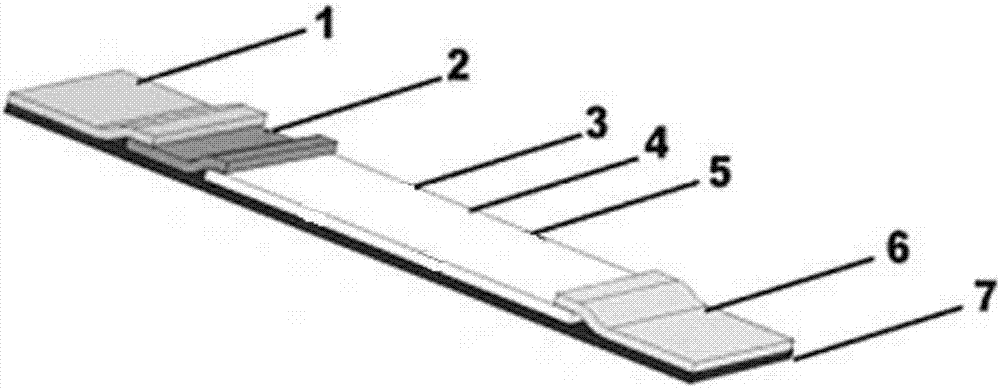
[0025] The first aspect of the present disclosure provides a method for preparing a human myeloperoxidase immunochromatographic test strip, characterized in that the immunochromatographic test strip includes a sample pad, a labeled antibody pad, an antibody pad, and an antibody pad sequentially connected. Coated with nitrocellulose membrane and absorbent pad; the labeled antibody pad contains 60-80 μg of colloidal gold or the first monoclonal antibody of fluorescently labeled human myeloperoxidase; the preparation method includes antibody-coated nitrocellulose The preparation of plain film and the assembly of immunochromatographic test strips, the preparation of described antibody-coated nitrocellulose membrane comprises the following steps:
[0026] S1. Mix the second monoclonal antibody solution of 1-2mg / mL human myeloperoxidase and 0.3-0.7% (W / V) 3-aminopropyltriethoxysilane phosphate buffer according to volume ratio 1: (4-6) mixed evenly to obtain 3-aminopropyltriethoxysil...
Embodiment 1
[0044] Take 20mL 40μg / mL colloidal gold solution in a 50mL clean centrifuge tube, after returning to room temperature, add 0.2mol / LK 2 CO 3 Adjust the pH to 8.0, and stir at a rate of 200 rpm under dark conditions. Add 10 μg of the primary monoclonal antibody to human myeloperoxidase into 0.5 ml of borate buffer and mix well, then slowly add it dropwise into the colloidal gold, and continue stirring for 20 min. Then slowly add 10% (W / V) bovine serum albumin dropwise to a final concentration of 1% (W / V), and continue stirring for 1 h. Stand overnight at 4°C to terminate the reaction. After the reaction, centrifuge at 15,000 rpm for 1 h at 4°C, and discard the supernatant. The precipitate is suspended in the bovine serum albumin solution of 1% (W / V) of original volume 1 / 10 to obtain the solution containing the first monoclonal antibody of human myeloperoxidase labeled with colloidal gold, and the solution is sprayed with gold The film dispenser evenly sprays on the glass cel...
Embodiment 2
[0048] The europium-containing polystyrene fluorescent microspheres were diluted to a concentration of 0.04mg / ml with 0.02M pH7.4 phosphate buffer solution, and then EDC (carbodiimide) with a final concentration of 2mM and NHS with a final concentration of 5mM were added (N-hydroxysuccinimide) was reacted at room temperature for 15 minutes, and the supernatant was removed by centrifugation at 15,000 rpm. Add 1.7 μL of the first monoclonal antibody of human myeloperoxidase with a concentration of 6 mg / mL to the precipitate, react at room temperature for 4 h, and then add 2% (W / V) glycine, 1% (W / V) bovine Serum albumin was treated with 0.02M pH 7.4 phosphate buffer for 30min, centrifuged to remove the supernatant, and then 1% (W / V) bovine serum albumin 0.02M pH 7.4 phosphate was added to the precipitate The buffer solution is suspended to obtain the first monoclonal antibody solution of human myeloperoxidase labeled with europium polystyrene fluorescent microspheres. The soluti...
PUM
| Property | Measurement | Unit |
|---|---|---|
| Sensitivity | aaaaa | aaaaa |
Abstract
Description
Claims
Application Information
 - R&D
- Intellectual Property
- Life Sciences
- Materials
- Tech Scout
- Unparalleled Data Quality
- Higher Quality Content
- 60% Fewer Hallucinations
Browse by: Latest US Patents, China's latest patents, Technical Efficacy Thesaurus, Application Domain, Technology Topic, Popular Technical Reports.
© 2025 PatSnap. All rights reserved.Legal|Privacy policy|Modern Slavery Act Transparency Statement|Sitemap|About US| Contact US: help@patsnap.com


