
Monomolecular fluorescence resonance energy transfer method based on photo-activation

A technology of fluorescence resonance energy and light activation, which is applied in the fields of fluorescence/phosphorescence, material excitation analysis, material analysis by optical means, etc.

Active Publication Date: 2017-07-04
TSINGHUA UNIV
View PDF4 Cites 1 Cited by
  • Summary
  • Abstract
  • Description
  • Claims
  • Application Information

AI Technical Summary

Problems solved by technology

Therefore, many studies using single-molecule FRET are carried out under conditions far below physiological concentrations, and whether the conclusions can truly reflect physiological conditions remains to be verified

Method used


Image

  • Monomolecular fluorescence resonance energy transfer method based on photo-activation
  • Monomolecular fluorescence resonance energy transfer method based on photo-activation

Examples


Embodiment 1

[0034] Example 1, Single-molecule fluorescence resonance energy transfer based on CAGE 552 photoactivatable dye.

[0035] Using the nucleic acid molecule labeled with CAGE 552 light-activated dye as a model, the observation of single-molecule fluorescence resonance energy transfer at a sample concentration of micromolar is realized.

[0036] The specific experiment is as follows:

[0037]1 mg of N-hydroxysuccinimide ester (NHS ester)-activated CAGE 552 photoactivatable dye was purchased from Abberior and dissolved in 181 μL of dimethyl sulfoxide so that the final concentration of the dye was 10 mM. CAGE 552 dye can be activated by 405nm laser and excited by 532nm laser. Purchase 1 mg of N-hydroxysuccinimide ester (NHSester)-activated Cyanine5 non-photoactivatable dye from Lumiprobe Company, and dissolve it in 162 μL of dimethyl sulfoxide, so that the final concentration of the dye is 10 mM. The biological sample is nucleic acid (DNA). A 100 μL 50 μM DNA single-stranded molec...

Embodiment 2

[0038] Example 2, single-molecule fluorescence resonance energy transfer based on CAGE FAM light-activated dye.

[0039] Using the nucleic acid molecule labeled with CAGE FAM light-activated dye as a model, the observation of single-molecule fluorescence resonance energy transfer at the sample concentration at the micromolar level is realized.

[0040] The specific experiment is as follows:

[0041] Purchase 1 mg of N-hydroxysuccinimide ester (NHS ester) activated CAGE FAM (CMNB-cagedcarboxyfluorescein, SE) photoactivatable dye from Invitroge, dissolve it in 103 μL of dimethyl sulfoxide, so that the final concentration of the dye is 10 mM . CAGE FAM dyes can be activated by 405nm laser and excited by 488nm laser. Purchase 1 mg of N-hydroxysuccinimide ester (NHS ester)-activated Cyanine5 non-photoactivatable dye from Lumiprobe, and dissolve it in 162 μL of dimethyl sulfoxide so that the final concentration of the dye is 10 mM. The biological sample is nucleic acid (DNA). A 1...


PUM


Abstract

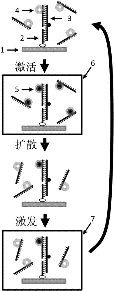
The invention discloses a monomolecular fluorescence resonance energy transfer method based on photo-activation, and belongs to the technical field of biological macromolecule imaging. A photo-activated fluorescent dye is used to label a biological sample taken as the donor. An activation light source with a wavelength of 200 to 450 nanometers is used to trigger the photo-activated fluorescent dye to carry out chemical reactions such as cis-trans isomerism reactions, electron transfer reactions, chemical bond breaking reactions, and the like. The photo-activated fluorescent dye is converted from a non-activated state that does not emit fluorescence into an activated state that can emit fluorescence. Then an excitation light source with a wavelength of 450 to 1200 nanometers is used to excite the fluorescent dye so as to emit fluorescence. When the distance between a donor and an acceptor labeled by a non-photo-activated fluorescence dye is in a range of 1-10 nanometers, the energy of the donor is transferred to the acceptor, namely fluorescence resonance energy transfer. The method has the advantages that in a conventional fluorescence resonance energy transfer technology, the concentration of a fluorescence sample should reach about 50 nM, while in the provided method the measurement of monomolecular fluorescence resonance energy transfer can be carried out under a micro-mole concentration, which is close to the physiological conditions.

Description

[0001] Technical field: [0002] The invention belongs to the field of biomacromolecule imaging, and more specifically relates to a new imaging technology and method that breaks through the fluorescence concentration barrier in the existing total internal reflection fluorescence microscopy technology. [0003] Background technique: [0004] Single-molecule fluorescence technology can observe the dynamic process of biomolecules in complex systems in real time, and reveal important information hidden in traditional ensemble average measurements, including the differences between biomolecules and the transient intermediate state during the reaction process. The above advantages of single-molecule fluorescence means have made it widely concerned and applied in recent years. Its rapid development has enabled many traditional biological problems to be solved with new experimental techniques (Sasmal D.K.et al., Nanoscale, 2016, 8, 19928- 19944). [0005] One of the most widely used s...

Claims


Application Information

Patent Timeline
IPC IPC(8): G01N21/64
CPCG01N21/6428
Inventor 陈春来王文娟彭思佳
Owner TSINGHUA UNIV
Learn More