
CT image spine and spinal dura mater automation detection method

An automatic detection and CT image technology, applied in the field of medical image processing, can solve the problems of time-consuming and laborious, increasing the workload of doctors, and achieve the effect of reducing the search range, improving efficiency, and being convenient and quick to use.

Inactive Publication Date: 2017-05-17
ZHEJIANG UNIV
View PDF3 Cites 6 Cited by
  • Summary
  • Abstract
  • Description
  • Claims
  • Application Information

AI Technical Summary

Problems solved by technology

[0003] The traditional judgment of the spine position based on the cross-sectional CT images of the spine is usually done manually, which is time-consuming and labor-intensive, which increases the workload of doctors

Method used


Image

  • CT image spine and spinal dura mater automation detection method
  • CT image spine and spinal dura mater automation detection method
  • CT image spine and spinal dura mater automation detection method

Examples


Embodiment Construction

[0035] Further illustrate the present invention below in conjunction with accompanying drawing.

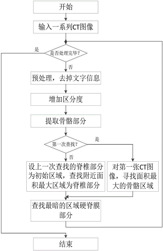
[0036] The CT image spine and dura mater automatic detection process of the present invention are as follows: figure 1 As shown, the process has the following steps in sequence:

[0037] 1) The user inputs a series of CT images A of the spine cross-section from top to bottom;

[0038] 2) For the i-th processing, take the i-th image A i , image A i Perform preprocessing to remove the text information in the CT image (such as Figure 4 Middle a area), get the image B with the text information removed i ;

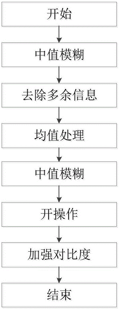
[0039] 3) For image B i Perform processing to increase the distinction between bones and other parts, and get image C i ;

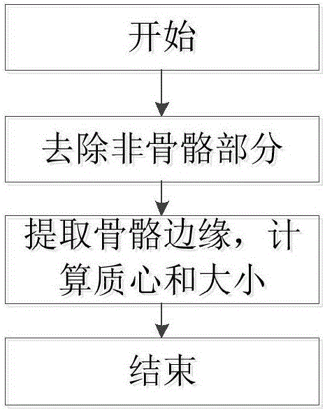
[0040] 4) Extract the bone area, find the centroid and size of each area, and get the image D i ;

[0041] 5) Filter out the board part during detection (such as Figure 4 Middle b area). By calculating the covariance of the edge of the bone area ...


PUM


Abstract

The invention discloses a CT image spine and spinal dura mater automation detection method. The method is characterized by using a series of input spinal cross sections to scan a CT image and automatically carrying out detection; filtering redundant information and extracting a portion where bones are located; and automatically detecting positions of a spine and a spinal dura mater in each image. The method is convenient and fast. Extra operation is not needed and a complicated and dull processing process needed by traditional manual determination can be avoided. The method is suitable for a series of inputs. During each time of detection, a previous detection result is taken as an initial result. And searching is performed near the initial result so that a searching range is reduced and efficiency is greatly increased.

Description

technical field [0001] The invention relates to the technical field of medical image processing technology, in particular to spine and dura mater detection and auxiliary diagnosis of medical images, in particular to an automatic detection method for spine and dura mater in CT images. Background technique [0002] With the rapid development of medical technology, more and more CT imaging technology is used in various fields, and CT imaging technology has also been widely used in the medical diagnosis of the spine. The detection of the spine is usually performed when diagnosing diseases such as intervertebral disc disease, spinal bone disease, and spinal injury. Due to the large number of patients who need to use CT to assist in the diagnosis of the spine, and each patient also has multiple CT images that need to be diagnosed, how to quickly and accurately judge the spine and related parts of the spine from a large number of CT images has become an urgent problem to be solved....

Claims


Application Information

Patent Timeline
IPC IPC(8): G06T7/00G06T7/13G06T7/62G06T7/66
CPCG06T7/0012G06T2207/10081G06T2207/30012
Inventor 于金辉吴凯琳张秉炜
Owner ZHEJIANG UNIV
Learn More