Terahertz wave attenuation total reflection imaging-based cerebral trauma tissue detection device

A technology of attenuated total reflection and terahertz waves, applied in diagnostic recording/measurement, medical science, sensors, etc., can solve problems such as weak anti-interference ability, difficulty in ensuring accuracy, surviving brain tissue, wounded, dead and disabled, etc., to achieve safety The effect of high accuracy, no interference fringes, and avoidance of destructive analysis

Pending Publication Date: 2017-04-26
TIANJIN UNIV
View PDF6 Cites 12 Cited by
  • Summary
  • Abstract
  • Description
  • Claims
  • Application Information

AI Technical Summary

Problems solved by technology

However, at present, the degree of brain tissue damage mainly depends on the empirical judgment of the operator, and it is difficult to guarantee the accuracy, which is not conducive to the treatment of high-throughput wounded patients.
In actual intracranial debridement emergency surgery, the boundary between inactivated brain tissue and surviving brain tissue is not clear and the boundary changes dynamically with the development of the injury and the progress of the operation. May cause massive casualties
[0003] At present, the methods that can be used to detect brain functional areas and inactivated brain tissue include: intraoperative electrophysiology, CT, MRI, magnetoencephalography and other detection methods used alone or in combination, but the above technologies have many limitations: ① Accurate Insufficient accuracy, intraoperative electrophysiological technology determines the range of functional areas through limited detection points, and the spatial and temporal resolution is poor. Although nuclear magnetic resonance and magnetoencephalography can achieve high spatial and temporal resolution, the equipment is bulky and The intraoperative implementation is difficult and the detection cost is expensive; ②The feasibility is not strong. First, the above-mentioned techniques require a high-demand anesthesia system for the detection of inactivated brain tissue, which is difficult to carry out in lower-level hospitals. Second, the magnetic examination system will cause damage to the brain tissue after trauma. ③The anti-interference ability is weak, and craniocerebral trauma is usually accompanied by foreign body residues in the brain. These residues will cause artifacts in the above inspection equipment, and the inspection results can hardly be used for treatment. Provide a reliable reference
[0005] The existing terahertz transmission imaging technology has extremely strict requirements on the thickness of fresh biological tissue samples, only tens of microns, which requires special sample preparation and water retention methods, which increases the production cost and time cost of samples
And the thickness is close to the THz wave measurement wavelength, which is easy to produce interference fringes

Method used


Image

  • Terahertz wave attenuation total reflection imaging-based cerebral trauma tissue detection device
  • Terahertz wave attenuation total reflection imaging-based cerebral trauma tissue detection device

Examples


Embodiment 1

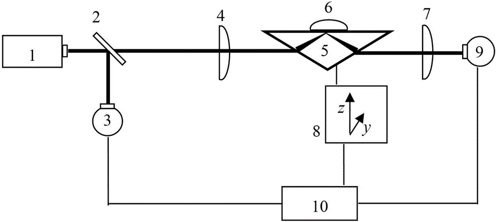
[0037] A brain trauma tissue detection device based on terahertz wave ATR imaging, see figure 1 , the detection device includes: a terahertz source 1, a terahertz beam splitter 2, a first terahertz aspheric lens 4 and a second terahertz aspheric lens 7, a total reflection prism 5, a biological sample to be measured 6, a two-dimensional moving scanning Platform 8 , first terahertz wave detector 3 , second terahertz wave detector 9 and computer control system 10 .

[0038] The terahertz source 1 emits terahertz waves with a frequency range of 0.1THz-30THz. The terahertz waves emitted by the terahertz source are incident on the terahertz beamsplitter 2, and the terahertz beamsplitter 2 is adjusted to form a certain angle with the incident terahertz waves. (10—80°), so that the incident terahertz wave is divided into a transmission terahertz wave and a reflected terahertz wave with a certain light intensity, and the terahertz wave reflected by the terahertz beam splitter 2 is used...

Embodiment 2

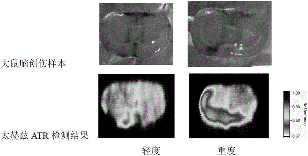
[0052] Combine below figure 2 , the feasibility verification of the device in Example 1 is carried out, see the following description for details:

[0053] In the embodiment of the present invention, the detection of brain trauma in mice is taken as an example, and a terahertz source with a frequency of 2.52 THz is used. First, two fresh rat brain tissue samples with different degrees of trauma were prepared by medical means. In order to make the biological sample 6 to be tested closely contact with the upper surface of the total reflection prism 5, the surface to be tested was repaired and flattened with a cryostat, and then the brain tissue Placed on the total reflection prism 5, use the computer control system 10 to control the movement of the total reflection prism 5 to realize point-by-point scanning, and collect each point of the rat brain tissue through the first terahertz detector 3 and the second terahertz detector 9 respectively Corresponding to the two light inten...


PUM


Abstract

The present invention discloses a terahertz wave attenuation total reflection imaging-based cerebral trauma tissue detection device. Terahertz waves emitted by a terahertz source enters a terahertz beam splitter which forms a certain angle with the terahertz waves; one part of the terahertz waves is reflected by the terahertz beam splitter, and the reflected terahertz waves enter a first terahertz detector which is connected with the input end of a computer control system; terahertz waves transmitted by the terahertz beam splitter are used as incident light for brain tissue sample detection, sequentially pass through a first terahertz aspherical lens, a total-reflection prism used for accommodating a brain tissue sample, and a second terahertz aspherical lens, and are finally detected by a second terahertz detector; the total-reflection prism is fixed on a two-dimensional mobile platform; and the computer control system receives data received by the first terahertz detector and the second terahertz detector and the moving information of the two-dimensional mobile scanning platform. With the terahertz wave attenuation total reflection imaging-based cerebral trauma tissue detection device of the invention adopted, the imaging of a brain trauma tissue is realized, and the location of the brain trauma tissue and the size of a lesion can be accurately embodied.

Description

technical field [0001] The invention relates to the field of brain trauma tissue detection devices, in particular to a brain trauma tissue detection device based on terahertz wave attenuated total reflection (ATR) imaging. Background technique [0002] Traumatic brain trauma is a major problem that threatens human life. In normal times, craniocerebral injury is the most important part of trauma, with a high rate of disability and mortality. Precise removal of inactivated brain tissue at an early stage and effective protection of brain functional areas are the keys to improving the success rate of traumatic brain injury treatment. However, at present, the degree of brain tissue damage mainly depends on the empirical judgment of the operator, which is difficult to guarantee accuracy and is not conducive to the treatment of high-throughput wounded patients. In actual intracranial debridement emergency surgery, the boundary between inactivated brain tissue and surviving brain ...

Claims


Application Information

Patent Timeline
IPC IPC(8): A61B5/00
CPCA61B5/00
Inventor 王与烨徐德刚陈图南冯华刘宏翔赵恒立姚建铨
Owner TIANJIN UNIV
Why Eureka
Try Eureka