
Method for segmenting liver medical images on basis of shape prior

A medical image and liver technology, applied in the field of medical image processing, to achieve a stable and effective liver shape prior and remove noise interference

Active Publication Date: 2017-01-25
HEBEI UNIVERSITY
View PDF5 Cites 1 Cited by
  • Summary
  • Abstract
  • Description
  • Claims
  • Application Information

AI Technical Summary

Problems solved by technology

Conventional methods in the field of medical image segmentation are active shape models and statistical shape models, however, modeling the shape of the liver in liver cancer patients is more challenging than modeling the shape of other organs such as prostate, kidney, and bone
The shape of the liver in different patients is ever-changing, which cannot be accurately described by a parameter probability distribution

Method used


Image

  • Method for segmenting liver medical images on basis of shape prior
  • Method for segmenting liver medical images on basis of shape prior
  • Method for segmenting liver medical images on basis of shape prior

Examples


Embodiment Construction

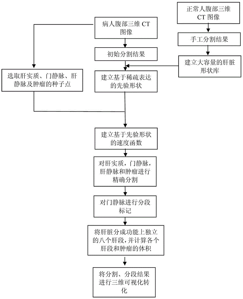
[0035] like figure 1 As shown, the realization method of the computer-aided liver transplant surgery planning system is:

[0036] The first step is to establish a large-capacity library of normal human liver shapes. The steps are as follows: extensively collect clinical data, and manually segment livers from at least 300 cases of three-dimensional abdominal CT images by clinical experts as the liver gold standard of corresponding individuals. The surface mesh on the gold standard is extracted and sampled to obtain a mesh surface composed of a series of marked points and triangle patches, thereby establishing a normal liver shape library.

[0037] In the second step, the region growing algorithm is used to obtain the initial segmentation result of the liver cancer patient or the donor liver to be operated, and the segmentation result is expressed as a sparse linear combination expression of the shapes in the shape library. The result of the sparse combination expression serve...


PUM


Abstract

The invention relates to a method for segmenting liver medical images on the basis of shape prior. Speed functions are defined by the aid of prior shapes, the arrival time of seed points which proceed and arrive at unclassified points is computed, and the medical images of livers and internal tissues of the livers are segmented according to the arrival time so as to obtain liver parenchyma, portal veins, liver veins and tumor. The method includes segmenting procedures of computing the speed functions combined with the prior shapes; carrying out classification on the basis of the arrival time. Each unclassified point is classified as a category with the shortest arrival time, so that segmentation results can be obtained. The method has the advantages that the shortcoming of interference of singular points and noise can be overcome for specific patients, and the method has important significance on reserving local details.

Description

[0001] This application is a divisional application of "A Realization Method of a Computer-Aided Liver Transplant Operation Planning System". The filing date of the original application: 2012.11.30, the application number: 201210505148.1, and the name of the invention is: A Computer-Aided Liver Transplant Operation Planning System implementation method. technical field [0002] The invention relates to a medical image processing technology, in particular to a method for segmenting liver medical images based on shape prior. Background technique [0003] In liver transplantation, accurate knowledge of patient-specific liver anatomy plays a decisive role in formulating surgical strategies. The volume of the transplanted liver should be sufficient to maintain the life needs of the recipient, while the remaining liver volume should be kept as large as possible to reduce damage to the liver donor. At the same time, since the anatomical structures of blood vessels and tumors in th...

Claims


Application Information

Patent Timeline
Patent Type & Authority Applications(China)
IPC IPC(8): G06T7/10
CPCG06T2207/30056G06T2207/30096
Inventor 董斌王国泰王洪瑞顾力栩
Owner HEBEI UNIVERSITY
Learn More