
Angiography method applied to optical coherence tomography and OCT system

An optical coherence tomography and vascular imaging technology, which is applied in the field of OCT systems, can solve the problems of eye movement sensitivity, insensitivity, and poor imaging, and achieve the effects of reducing sensitivity, improving sensitivity, and reducing noise

Active Publication Date: 2016-11-30
WENZHOU MEDICAL UNIV
View PDF6 Cites 12 Cited by
  • Summary
  • Abstract
  • Description
  • Claims
  • Application Information

AI Technical Summary

Problems solved by technology

In biological tissues with high dispersion, such as the scleral tissue in front of the eye, there are still some problems with the SSADA and CM methods. The SSADA method is not sensitive to imaging of tiny blood vessels in high dispersion tissues, and cannot be well imaged. Sensitivity, but it is easily sensitive to eye movement, resulting in obvious noise
Therefore, OCT angiography still has certain difficulties in high dispersion biological tissues, such as angiography of the anterior segment.

Method used


Image

  • Angiography method applied to optical coherence tomography and OCT system
  • Angiography method applied to optical coherence tomography and OCT system
  • Angiography method applied to optical coherence tomography and OCT system

Examples


Embodiment Construction

[0028] The present invention can be better described below in conjunction with the accompanying drawings and specific embodiments.

[0029] Such as figure 1 As shown, the light emitted by the frequency-sweeping light source 1 passes through the first fiber coupler 3 with an ratio of 80:20, 20% of the light enters the sample arm 12 , and 80% of the light enters the reference arm 11 . Both the sample arm 12 and the reference arm 11 have polarization controllers 6 to adjust the polarization. The light entering the sample arm 12 is irradiated on the anterior segment of the human eye through the collimating mirror and the focusing lens, and three-dimensional data acquisition is realized through the swing of the vibrating mirror 9 . 80% of the light enters the reference arm and returns through dispersion compensation 7 and mirror 8 . The light returning from the reference arm and the sample arm enters a 50:50 second fiber coupler and acquires the interference signal at the balance...


PUM


Abstract

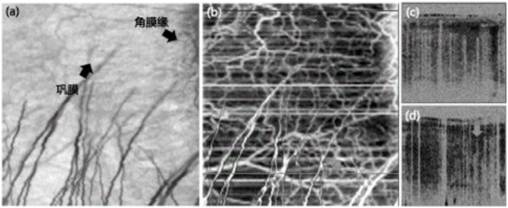
The invention relates to an angiography method applied to optical coherence tomography and an OCT system. Based on the frequency division thought, OCT interference fringes are decomposed into multiple wave number bands, and noise generated when shot tissue moves is lowered. After intensity images obtained after frequency division are acquired, an improved CM method is further adopted, the change degree of the intensity, calculated through the Pearson correlation coefficient, between adjacent continuously scanned cross-sectional images is calculated, and signals of blood vessels are enhanced. In combination with information of points adjacent to detection points, sensitivity to blood vessel detection is improved, sensitivity to eye movement is reduced, and the method is suitable for angiography of biological tissue with high scattering. The method can be used for angiography of anterior segment sclera or irises or other tissue in ophthalmology and can be applied to microvascular imaging of other portions of the human body.

Description

technical field [0001] The invention relates to an angiography method and an OCT system applied to optical coherence tomography. Background technique [0002] Microvascular morphology imaging has important applications in medical imaging. The evaluation of microvascular morphology is of great value to the diagnosis, monitoring and treatment evaluation of diseases, especially vascular diseases. Ex vivo and invasive imaging methods can obtain high-resolution three-dimensional morphological imaging, but at present, such imaging technologies are slow and have certain damage to the human body, which limits their application in clinical diagnosis. Therefore, optical-based imaging methods are non-invasive It is an important means to observe the state of mind of blood vessels in the body, and has important application value in clinical medicine. [0003] At present, there are many optical imaging technologies used for imaging the vascular morphology of biological tissues. Fluores...

Claims


Application Information

Patent Timeline
IPC IPC(8): A61B5/00A61B5/02A61B3/10G06T15/00
CPCA61B3/102A61B5/0066A61B5/02007A61B5/72G06T15/00G06T2210/41
Inventor 黄胜海沈梅晓朱德喜王媛媛吕帆
Owner WENZHOU MEDICAL UNIV
Learn More