


Quick Research
Generate reliable direction feasibility study reports for your R&D in just a few steps.

Technical Q&A
Discover and master advanced knowledge NOW. Basics, ideas, possibilities, all at once.

Find Solutions
As an expert in R&D theories, this can generate solutions to your technical problems instantly.

Evaluate Feasibility
Analyze your overall solution with one click, know your potential R&D risks in advance.

Monitor Landscape
Get weekly tech updates, stay abreast of the latest tech innovations and key insights.
Method for quantitatively detecting biological marker by utilizing glucose meter
A biological marker and quantitative technology, applied in biological testing, material inspection products, etc., can solve the problems of high cost, long time, not suitable for rapid detection and identification of biological markers, etc., and achieve low cost and short time consuming. Effect
- Summary
- Abstract
- Description
- Claims
- Application Information
AI Technical Summary
Problems solved by technology
Method used
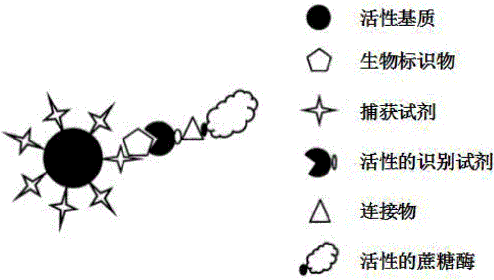
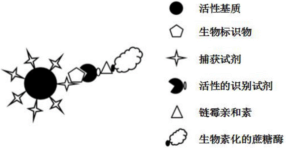
Image

Examples
preparation example Construction
[0084] In the present invention, the preparation method of the recognition reagent linked with sucrase in the step B) comprises the following steps:
[0085] The sucrase and the coupling agent are mixed and connected for 30 to 60 minutes to obtain the coupling agent-sucrase complex;
[0086] Mix the thiol-modified recognition reagent, PB buffer and tris(2-carboxyethyl)phosphine, and let stand for 30-60 minutes to obtain the activated recognition reagent;
[0087] The coupling agent-sucrase complex is mixed with the activated recognition reagent and left to stand for 24-48 hours to obtain the recognition reagent linked with the sucrase.
[0088] In the present invention, the reaction temperature for the mixing and connection of the sucrase and the coupling agent is preferably 20-30° C., and the mixing and connection is preferably performed at a rotation speed of 20-30 r / min.
[0089] In the present invention, the preparation method of the recognition reagent connected with suc...
Embodiment 1
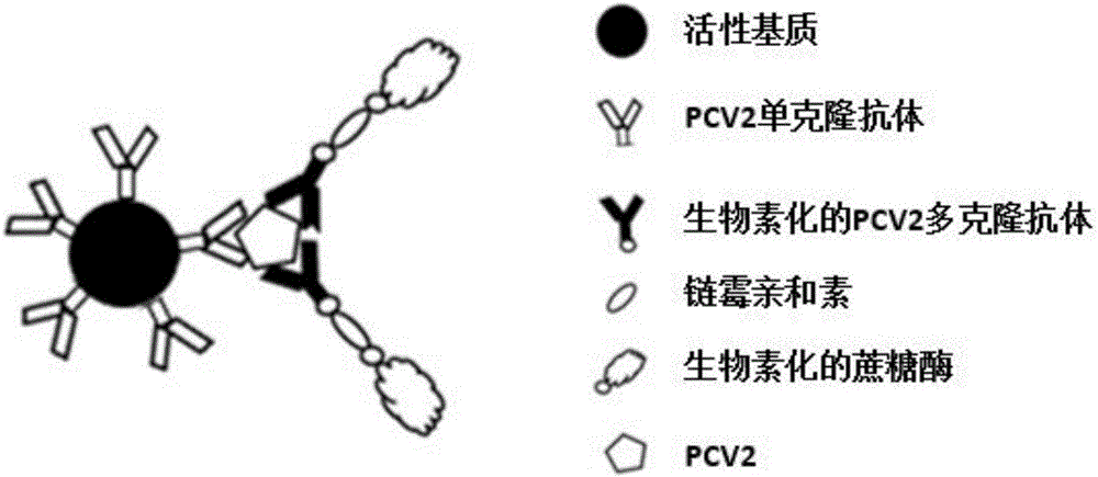
[0095] Link 100 μL of activated carboxyl magnetic beads with porcine circovirus type 2 PCV2 monoclonal antibody to obtain carboxyl magnetic beads with monoclonal antibody, then perform magnetic separation and wash 4 times with Buffer A solution. After magnetic separation, disperse the carboxyl magnetic bead precipitate in 100 μL of Buffer A solution containing PCV2 capsid protein—cap protein, and then mix it for 2 hours with a rotary mixer at 25°C and 25 r / min, and then perform magnetic separation of carboxyl magnetic beads. Beads were washed 3 times with Buffer A solution. Add 100 μL 1mg / L biotinylated PCV2 polyclonal antibody (dissolved in Buffer A solution) to the precipitation of carboxyl magnetic beads, then mix at 28°C for 45 minutes, magnetically separate carboxyl magnetic beads, wash 4 times with Buffer A solution, and carboxyl magnetic The bead precipitate was dispersed in 80 μL of 2 μM streptavidin (dissolved in Buffer A solution), and mixed for 50 min. Magnetically...
Embodiment 2
[0097] Link 60 μL of activated carboxyl magnetic beads to porcine circovirus type 2 PCV2 monoclonal antibody to obtain activated carboxyl magnetic beads, then perform magnetic separation and wash 3 times with Buffer A solution. After magnetic separation, disperse the carboxyl magnetic bead pellet in 100 μL of Buffer A solution (containing 25% pig serum) containing PCV2 capsid protein—cap protein, and then mix it with a rotary mixer at 28°C and 24r / min. 2h, then carry out magnetic separation of carboxyl magnetic beads, wash 3 times with Buffer A solution. Add 55 μL 1mg / L biotinylated PCV2 polyclonal antibody (dissolved in Buffer A solution) to the precipitation of carboxyl magnetic beads, then mix at 28°C for 55 min, magnetically separate carboxyl magnetic beads, wash twice with Buffer A solution, and remove the magnetic beads The precipitate was dispersed in 60 μL of 2 μM streptavidin (dissolved in Buffer A solution), and mixed for 45 minutes. Magnetically separate the carbox...
PUM
 Abstract
Description
Claims
Application Information
 - R&D Engineer
- R&D Manager
- IP Professional
- Industry Leading Data Capabilities
- Powerful AI technology
- Patent DNA Extraction
Browse by: Latest US Patents, China's latest patents, Technical Efficacy Thesaurus, Application Domain, Technology Topic, Popular Technical Reports.
© 2024 PatSnap. All rights reserved.Legal|Privacy policy|Modern Slavery Act Transparency Statement|Sitemap|About US| Contact US: help@patsnap.com